
Amoebiasis in freshwater is a particular problem in salmonids and is reported worldwide. It is sometimes given the name nodular gill disease (NGD), based on the marked and grossly observable epithelial hyperplasia. In Chile the first report was in February 2017, in rainbow trout in the Araucanía region.

Different genera of freshwater amoebae have been described for different fish species. Affected species include Atlantic salmon (Salmo salar), Chinook salmon (Oncorhynchus tshawytscha), rainbow trout (Oncorhynchus mykiss), Coho salmon (Oncorhynchus kisutch), blue tilapia (Sarotherodon aureus), golden carp (Carassius auratus), catfish (Silurus glanis) and Arctic trout (Salvelinus alpinus), among others. Species of amoebae include Cochliopodium, Acanthamoeba, Hartmannella, Naegleria, Protacanthamoeba and Vannella.

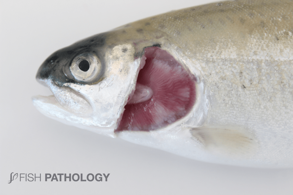
The clinical signs recorded in fish affected by nodular gill disease (NGD) are limited mainly to the gills; systemic involvement is uncommon except sometimes in severe cases for limited hepatic necrosis, associated with consequential hypoxaemia. Macroscopically, changes include diffuse pallor, excess mucus, and epithelial hyperplasia manifested as white nodules. Sometimes entire arches are completely obliterated due to hyperplastic epithelium.
Predisposing factors for the appearance of NGD are not fully understood, although poor water quality and elevated temperature can aggravate infections; so too can concurrent or preexisting infections such as bacterial gill disease. It is thought that the amoebae are initially attracted by the bacteria on the surface of the gills.
REFERENCES
- Ferguson, H.W., 2006, Systemic Pathology of Fish, London, UK, Scotian Press.
- Iva Dyková and Jiří Lom, Jill M. Schroeder-Diedrich, Gregory C. Booton and Thomas J. Byer, Acanthamoeba strains isolated from organs of freshwater fishes, Institute of Parasitology, Academy of Sciences of the Czech Republic, Branišovská 31, 370 05 České Budějovice, Czech Republic.
- K. Sawyer, Thomas & G. Hnath, John & F. Conrad, John. (1974). Thecamoeba hoffmani sp. n. (Amoebida: Thecamoebidae) from Gills of Fingerling Salmonid Fish. The Journal of parasitology. 60. 677-82. 10.2307/3278738.
- Tubbs L, Wybourne BA, Lumsden JS, Nodular gill Disease causing proliferative branchitis and mortality in Chinook salmon (Oncorhynchus tshawytscha) New Zealand Veterinary Journal, Volume 58, Issue 1, pp 59-61, Feb 2010.
