
Ichthyophthirius multifiliis is an opportunistic or ectocommensal parasite of skin and gills of freshwater fish. It is a common problem in farmed fish as well as aquarium species and in all water temperatures. It is a holotrichous ciliate, class Oligohymenophora, subclass Hymenostomata, order Hymenostomatida, suborder Ophryoglenina, family Ichthyophthiridae.
Among the various factors considered favourable to the parasite are poor water quality, high levels of decomposing organic matter, high stocking density, stress in handling, inadequate transport and low nutritional condition of the fish.
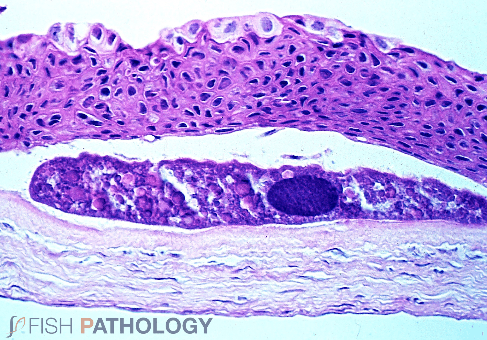
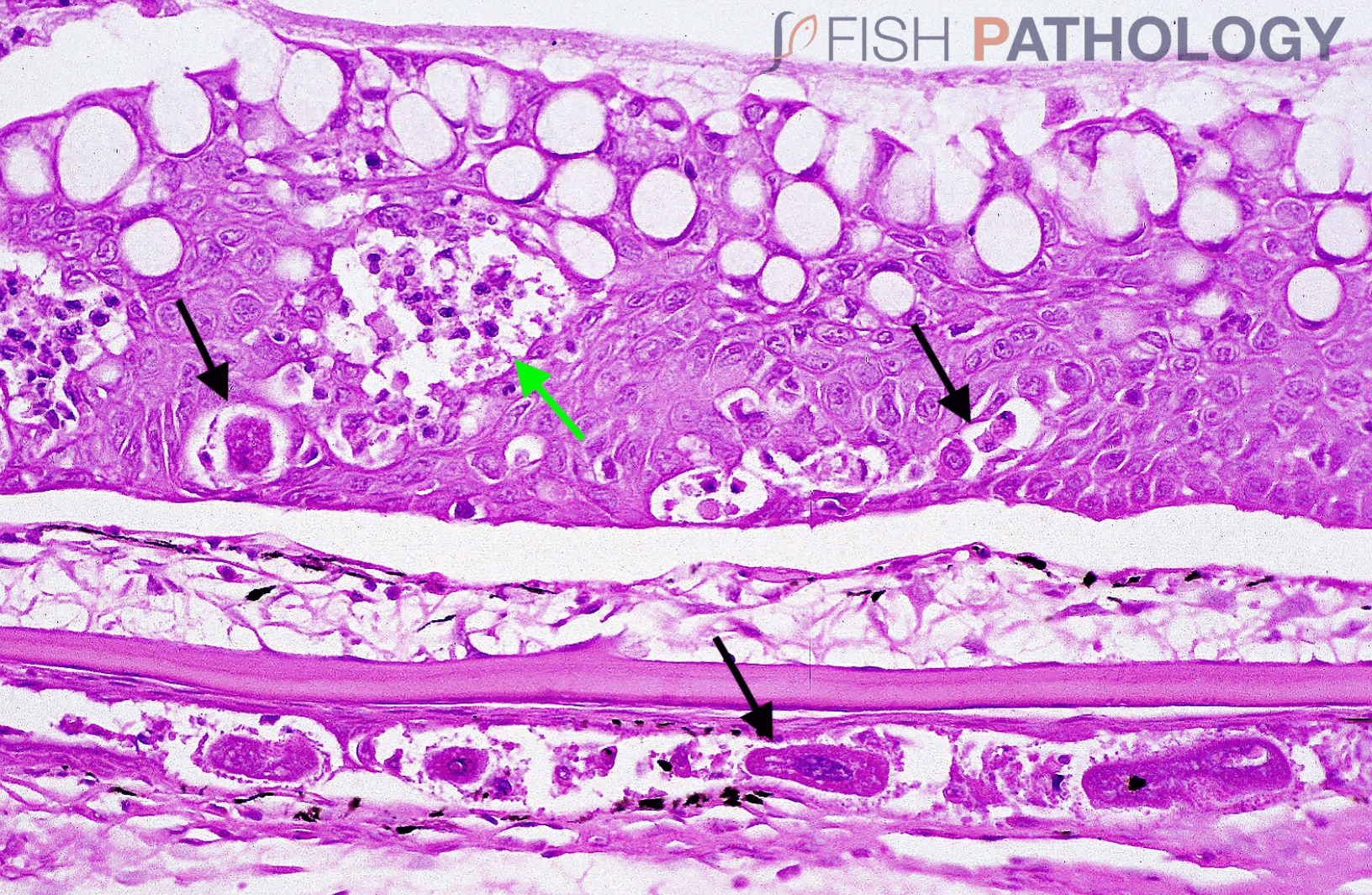
This disease is usually called “White Spot Disease”, since the parasite can be seen with the naked eye as one mm diameter white spots distributed over the body and fins when it is in the trophozoite stage, the feeding phase of the parasite. It should be noted that, despite appearances, the parasites actually sit within the epidermis, and not on the surface. This makes treatment more complicated, as a topical approach will not easily work.
Structurally, the entire surface of the organism is covered by motile cilia that are responsible for its motility in water, as well as its movement within the host tissue.
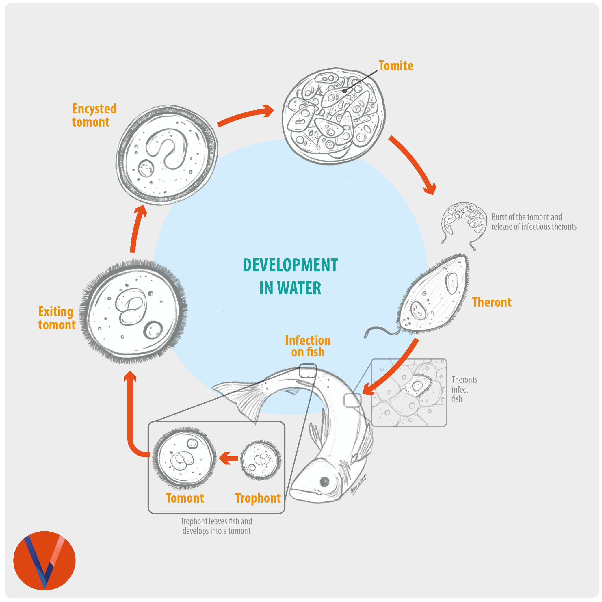
These parasites have a complicated life cycle that includes stages within the host as well as in the environment. The time taken for development within the fish is temperature-dependent; for example it requires 3–4 days at 22°C, up to 11 days at 15°C, and nearly 30 days at 10°C.
As soon as the theronts (infectious stage) begin to infect their host, the irritated fish can be seen shaking and rubbing their opercula and flanks against surfaces. The host cellular response is usually minimal but heavy infections of the gills can result in lamellar fusion leading to reduced respiratory function and electrolyte imbalance.
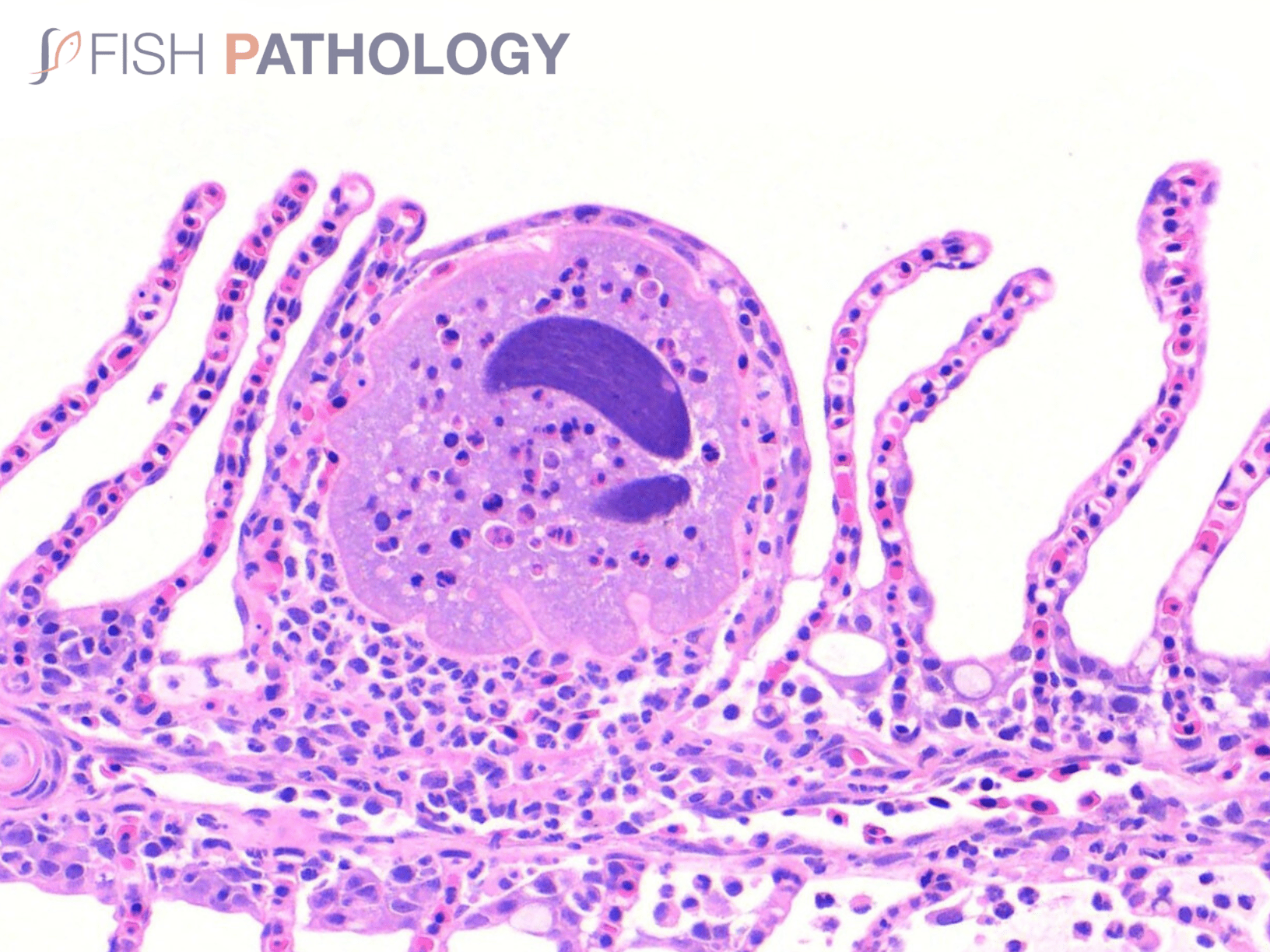
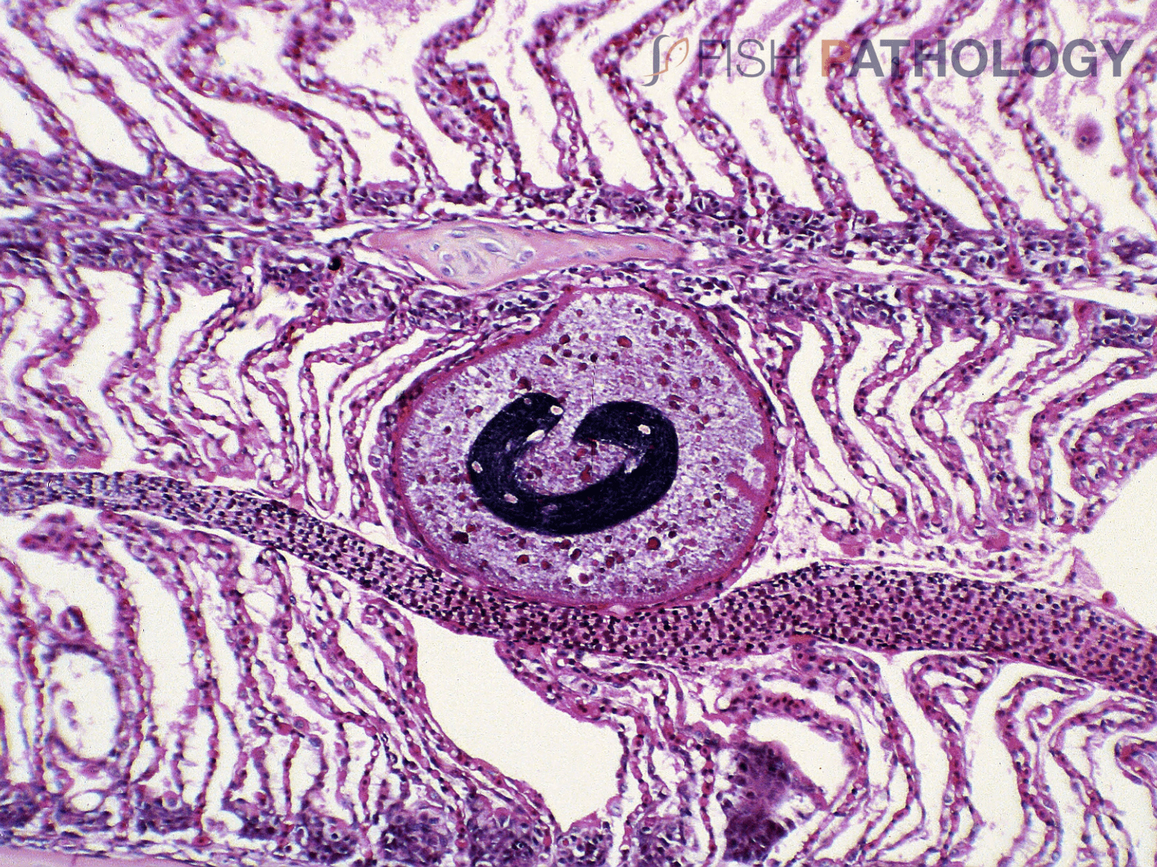
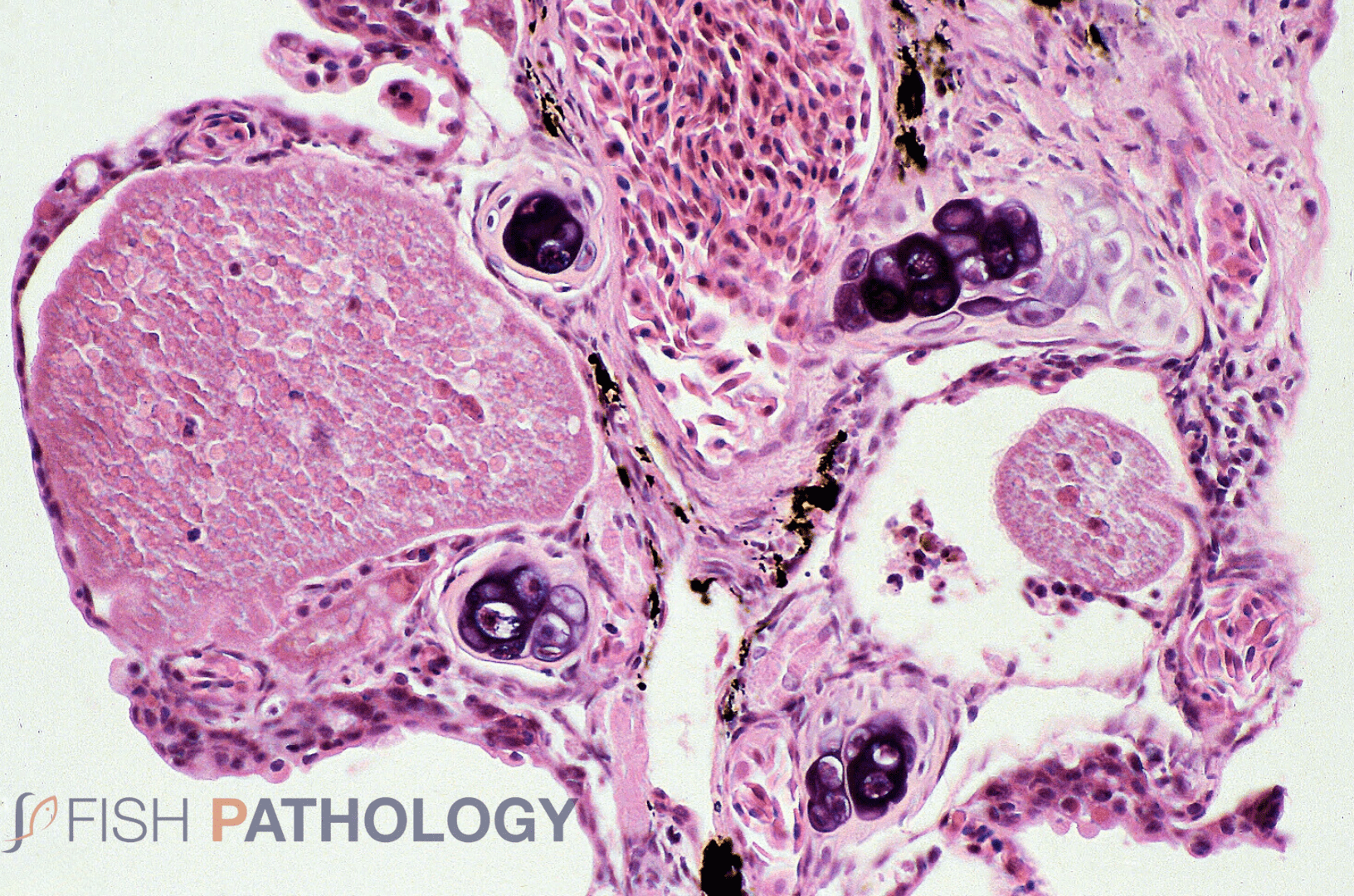
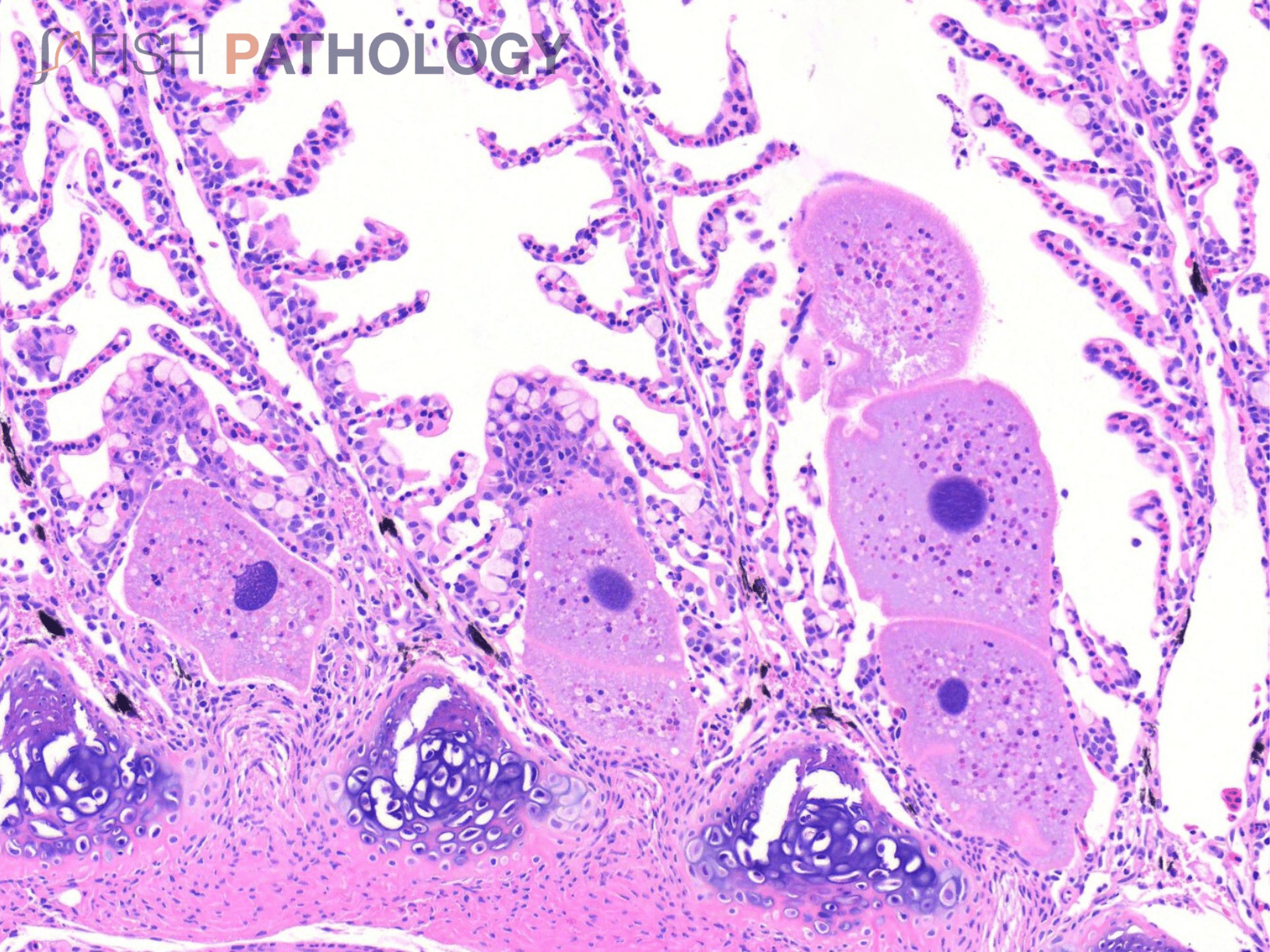
In histological sections of skin and gills, parasites are observed within the epithelium, usually sitting on the basement membrane. Structures from the parasite like the horseshoe-shaped macronucleus, cytoplasmic food vacuoles and superficial cilia are usually visible. When infections are intense, there may be hyperplasia and inflammatory cell infiltration, as well as necrosis and vacuolar degeneration of epithelial cells that can often be severe enough to cause clinical disease. This is especially true if the host is partially immune i.e. has had prior exposure. Once mature, the parasites erupt through the epithelium and encyst within the substrate to continue their life cycle. In heavy infections, large numbers of parasites erupting through the epidermis at the same time can have a severe impact on the host.
Images
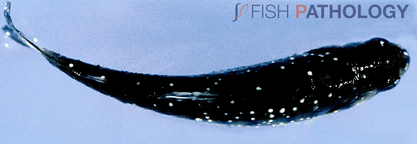
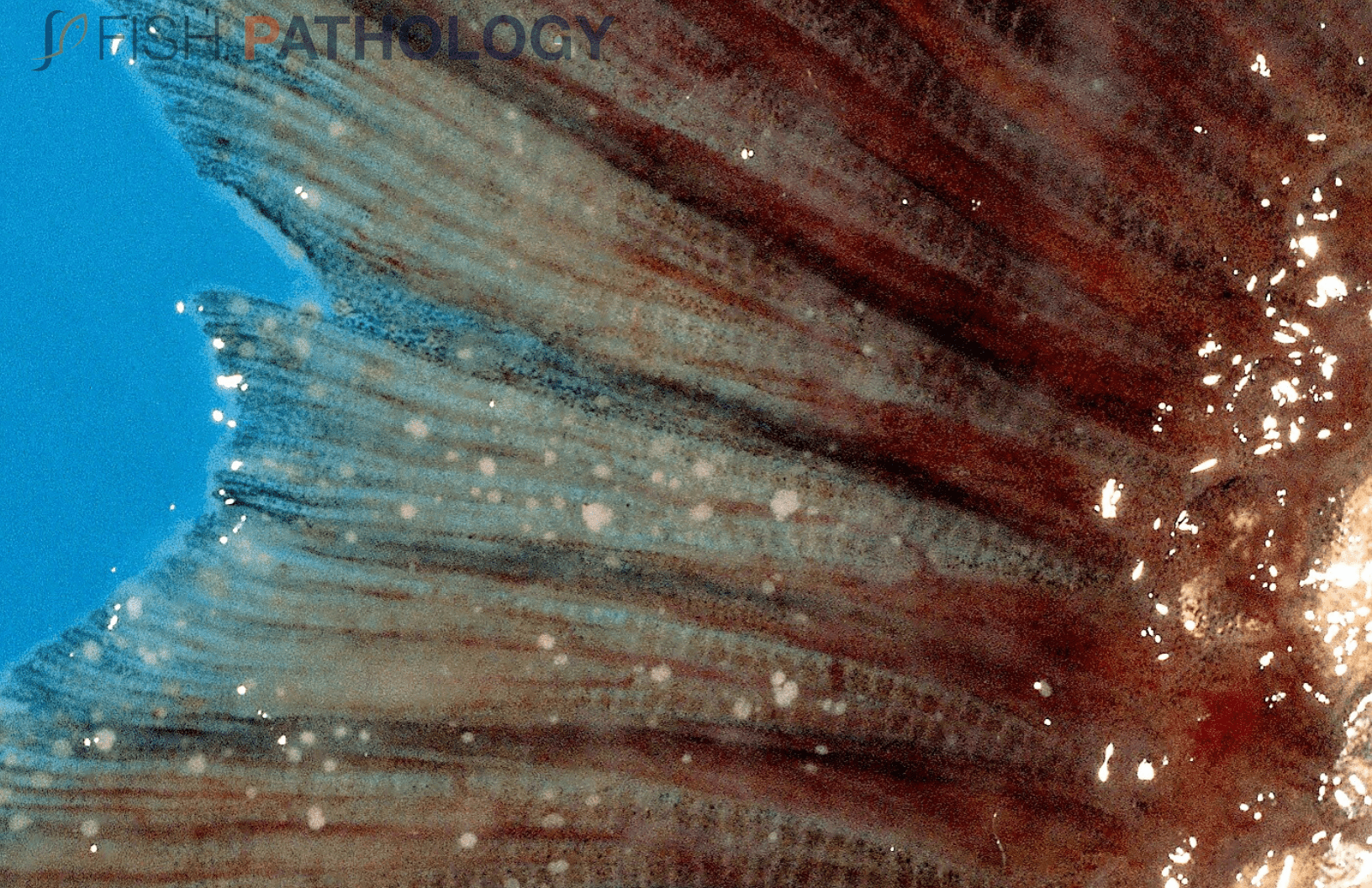
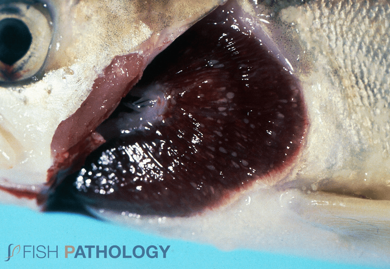
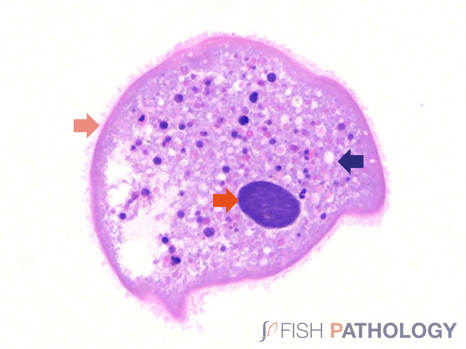
Figure 4. Histopathology in S. salar. Identification of the macronucleus (red arrow), feeding vacuoles (blue arrow) and cilia (orange arrow) in Ichthyophthirius multifiliis.
References
Casas, F. C., Ortiz, A. A., Sarabia, D. O., & Soriano, L. A. C. (1997). Infección por Aeromonas hydrophila e lchthyophthirius multifiliis en trucha (Oncorhynchus mykiss, Walbaum) y tilapia (Oreochromis aureus, L) de un centro de acopio de Morelos, México. Estudio patológico. Veterinaria México, 28 (1), 59-62.
Dickerson, H., & Clark, T. (1998). Ichthyophthirius multifiliis: a model of cutaneous infection and immunity in fishes. Immunological Reviews, 166(1), 377–384.
Ferguson, H. (2006). Systemic Pathology of Fish, A text and Atlas of Normal Tissues in Teleosts and their Responses in Disease, Second Edition, 55.
Gonzáles-Fernández, J. (2012). Parasitofauna en variedades del pez ornamental Carassius auratus y descripción del ciclo biológico de Ichthyophthirius multifiliis (Ciliatea, Ichthyophthiriidae), causante de mortalidades en un criadero de Lima, Perú, 2007. Neotropical Helminthology, vol. 6, nº 1, 85 – 95.
Jørgensen, L. von G. (2017). The fish parasite Ichthyophthirius multifiliis – Host immunology, vaccines and novel treatments. Fish & Shellfish Immunology, 67, 586–595.
Stoskopf, M. K. (2015). Biology and Management of Laboratory Fishes. Laboratory Animal Medicine, 1063–1086.
Woo, P., Buchmann, K. (2012). Fish Parasite, Pathobiology and Protection, 55 – 67.
Xiaoli, H., Senyue, L., Fengyuan, Z., Lin, L., Defang, C., Yangping, O., Yi, G., Yufan, Z., Gang, L., Shiyong, Y., Wei, L., Lizi, Y., Zhi, H. (2022). cMOS enhanced the mucosal immune function of skin and gill of goldfish (Carassius auratus Linnaeus) to improve the resistance to Ichthyophthirius multifiliis infection. Fish & Shellfish Immunology, 126, 1-11.
