
Tenacibaculosis is a serious bacterial disease affecting a great variety of marine fish, especially those species under culture conditions, causing necrotic lesions on the body. Gross pathological signs vary according to the species and age of fish involved. Characteristic clinical signs are ulcerative skin lesions, mouth erosion and ulceration, and fraying of fins and tail. In general, it is mainly a superficial infection, but some isolates are highly toxigenic, and systemic disease can therefore result, involving different internal organs.
Several species of Tenacibaculum can be involved, including T. dicentrarchi, T. finnmarkense (common in Norway), T. maritimum T. soleae, T. discolor, and T. gallaicum. T. maritimum is considered the main causative agent of Tenacibaculosis.
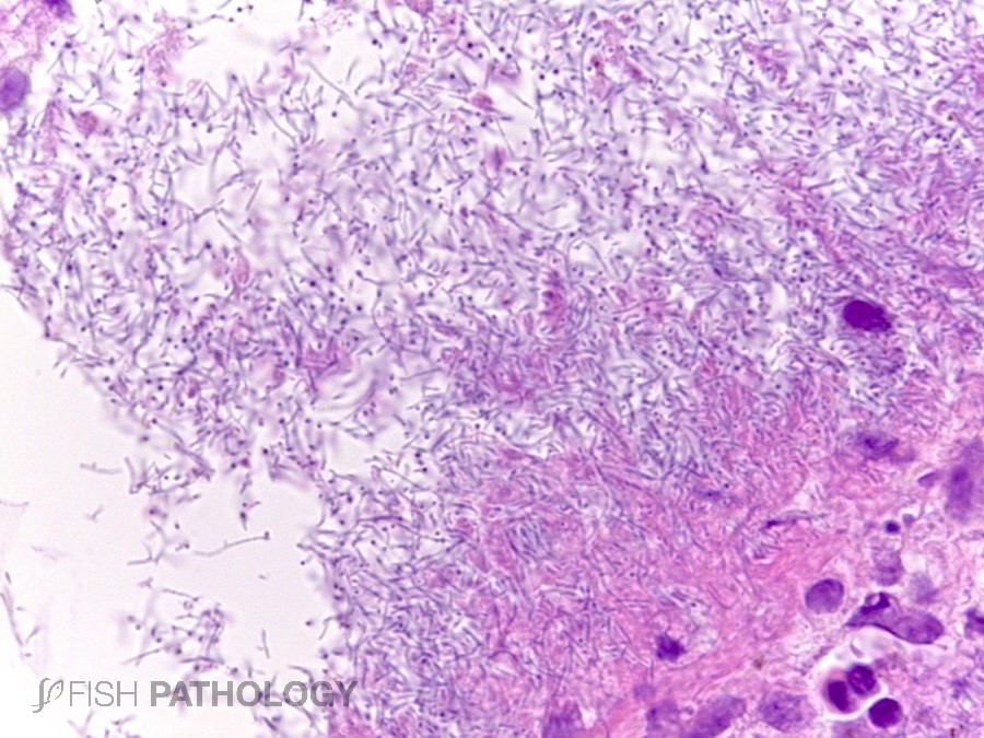
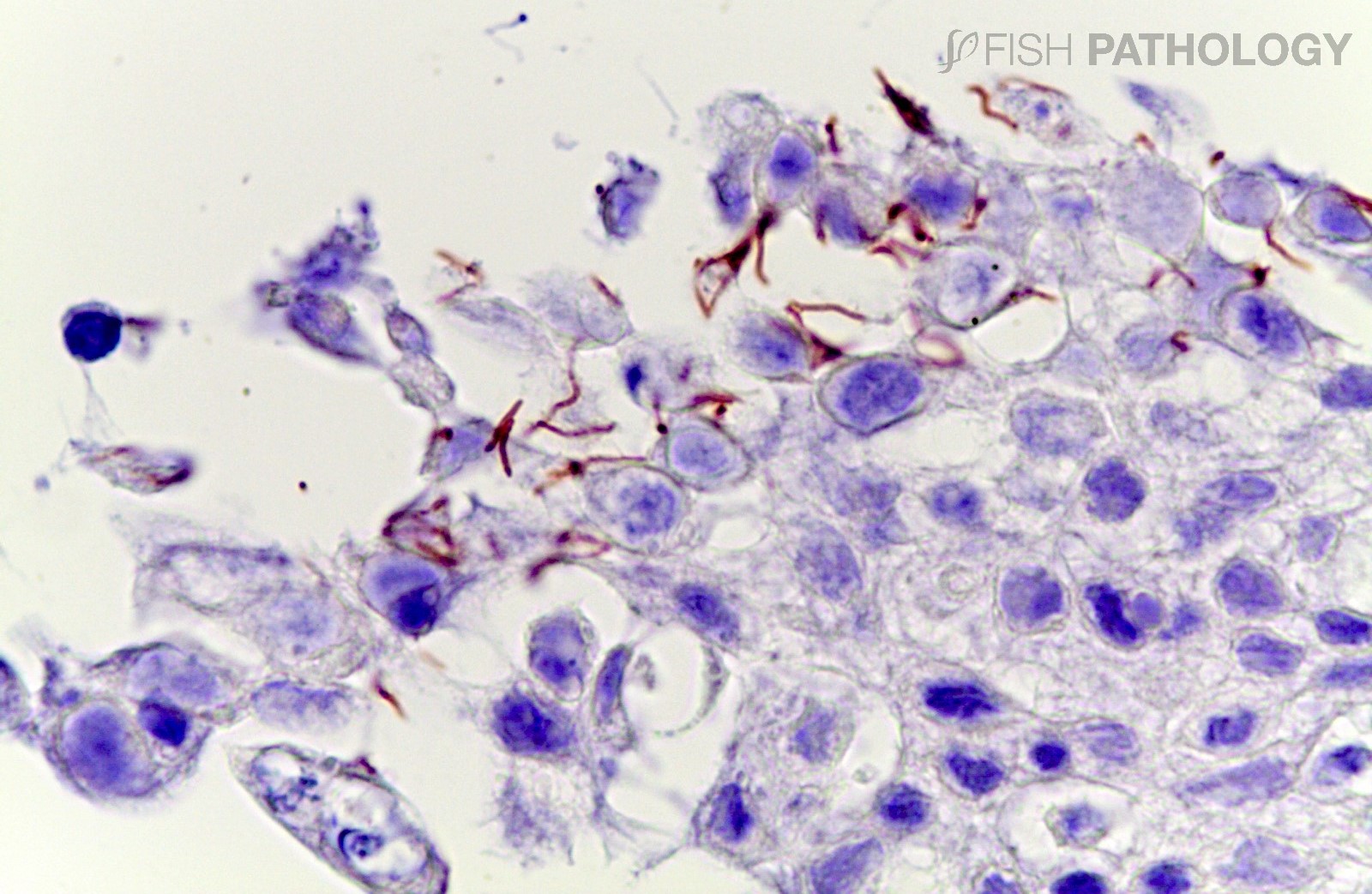
Dermal lesions typically include spongiosis, necrosis and ulceration in epidermis, and degeneration of collagen (necrobiosis) in dermis. Large clusters or mats of filamentous bacteria are usually seen, accompanied by a low-grade inflammatory reaction, mainly in dermis. In the healing process, fibrosis can be observed, with extensive mononuclear cell infiltration; scarring, however, does not occur.
Dermal lesions caused by T. dicentrarchi can include epidermal loss, with low-grade neutrophilic and lymphocytic infiltrations in dermis and hypodermis, along with congestion and multifocal haemorrhage; multifocal muscular degeneration and necrosis with haemorrhage can be seen in more advanced stages.
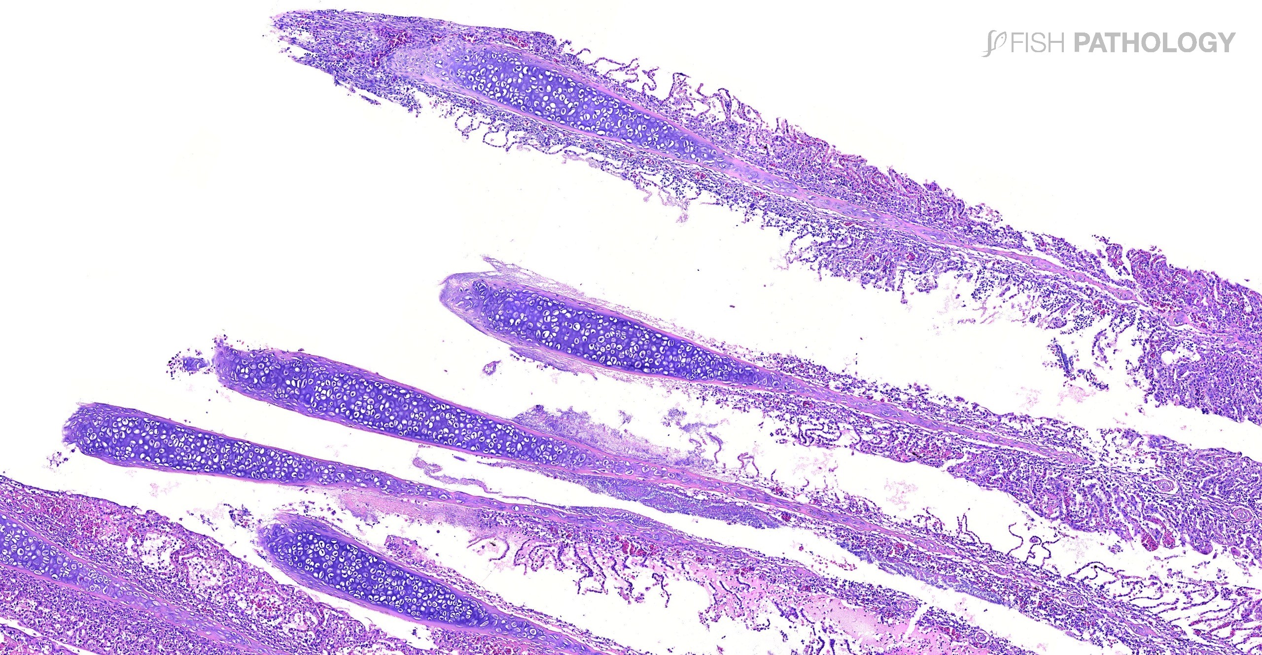
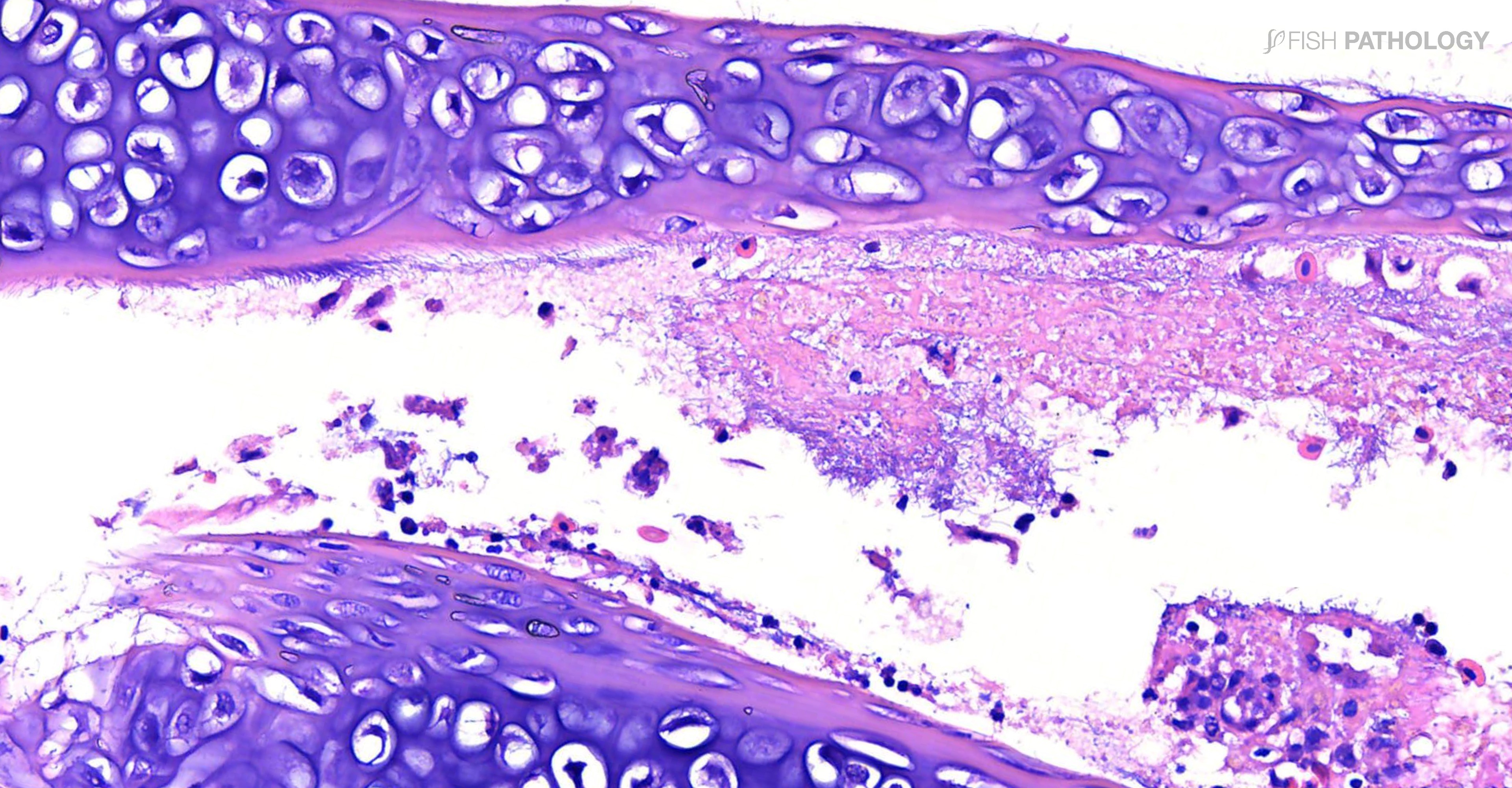
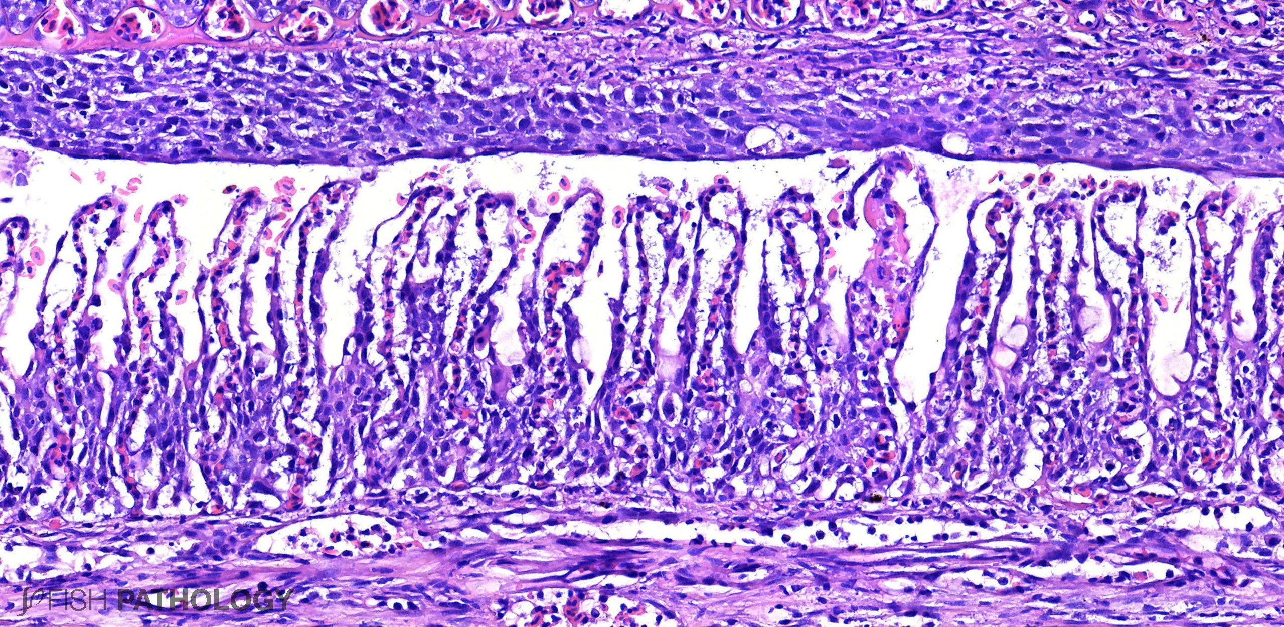
Gill lesions include focal to focally extensive areas of erosion and necrosis in branchial filaments, their cartilage, and in lamellae, with little or no accompanying inflammation. These lesions are usually associated with mats of filamentous bacteria overlaying necrotic tissue. Accompanying changes include epithelial lifting and sloughing, hypertrophy, telangiectasia, thrombosis, and occasionally an increase in the number of chloride cells.
In Atlantic salmon necrotising stomatitis is commonly seen. Glossitis and stomatitis can progress to necrotising cellulitis with osteitis/chondritis and may be severe enough to perforate the mandible. Teeth can also be involved, becoming loose and, in some cases, falling out. In severe cases, normal tissue structures are replaced by an amorphous mass comprising large amounts of bacteria and cellular debris. The reasons why T. maritimum targets the teeth and surrounding mucosa in this “mouth rot” are not fully understood. Possibilities include hard feed pellets damaging gingiva, and improper “cleaning” due to frequent/constant feeding practices. Regardless, teeth are a rich source of calcium, and this has been shown to promote the growth of T. maritimum.
Even seemingly inconsequential lesions in the mouth should not be overlooked as they can point to systemic involvement. While bacterial toxins may suppress host responses to some degree, typical systemic lesions may nevertheless include myocarditis and epicarditis, enteritis, multi-focal hepatitis, and pancreatitis, along with haemosiderosis in spleen and kidney – evidence of increased red cell breakdown.
REFERENCES
- Avendaño‐Herrera, R., Irgang, R., Sandoval, C., Moreno‐Lira, P., Houel, A., Duchaud, E., & Ilardi, P. (2016). Isolation, characterization, and virulence potential of Tenacibaculum dicentrarchi in salmonid cultures in Chile. Transboundary and emerging diseases, 63(2), 121-126.
- Ferguson, H.W. (2006). Systemic Pathology of Fish: a text and atlas of normal tissues in teleosts and their responses in disease. Second Edition, Scotian Press, London.
- Fernández-Álvarez, C., & Santos, Y. (2018). Identification and typing of fish pathogenic species of the genus Tenacibaculum. Applied microbiology and biotechnology, 102(23), 9973-9989.
- Frisch, K., Småge, S. B., Johansen, R., Duesund, H., Brevik, Ø. J., & Nylund, A. (2018). Pathology of experimentally induced mouthrot caused by Tenacibaculum maritimum in Atlantic salmon smolts. PLOS ONE, 13(11), e0206951.
- Gourzioti, E., Kolygas, M. N., Athanassopoulou, F., & Babili, V. (2016). Tenacibaculosis in aquaculture farmed marine fish. Journal of the Hellenic Veterinary Medical Society, 67(1), 21-32.
- Haridy, M., Hasheim, M., Abd El-Galil, M., Sakai, H., & Yanai, T. (2015). Pathological Findings of Tenacibaculum maritimus Infection in Black Damselfish, Neoglyphieodon melas and Picasso Triggerfish, Rhinecanthus assasi in Red Sea, Egypt. Veterinary Science & Technology, 6(2), 1.
- Lagadec E, Småge SB, Trösse C, Nylund A (2021). Phylogenetic analyses of Norwegian Tenacibaculum strains confirm high bacterial diversity and suggest circulation of ubiquitous virulent strains. PLOS ONE 16(10), e0259215.
- Nowlan, J. P., Lumsden, J. S., & Russell, S. (2020). Advancements in Characterizing Tenacibaculum Infections in Canada. Pathogens, 9(12), 1029.
- Nowlan, J. P., Lumsden, J. S., Britney, S. R. & Russell, S. (2021). Experimental Induction of Tenacibaculosis in Atlantic Salmon (Salmo salar L.) Using Tenacibaculum maritimum, T. dicentrarchi, and T. finnmarkense. Pathogens, 10(11), 1439.
- Van Gelderen, R., Carson, J., & Nowak, B. (2009). Effect of extracellular products of Tenacibaculum maritimum in Atlantic salmon, Salmo salar L. Journal of fish diseases, 32(8), 727-731.
