
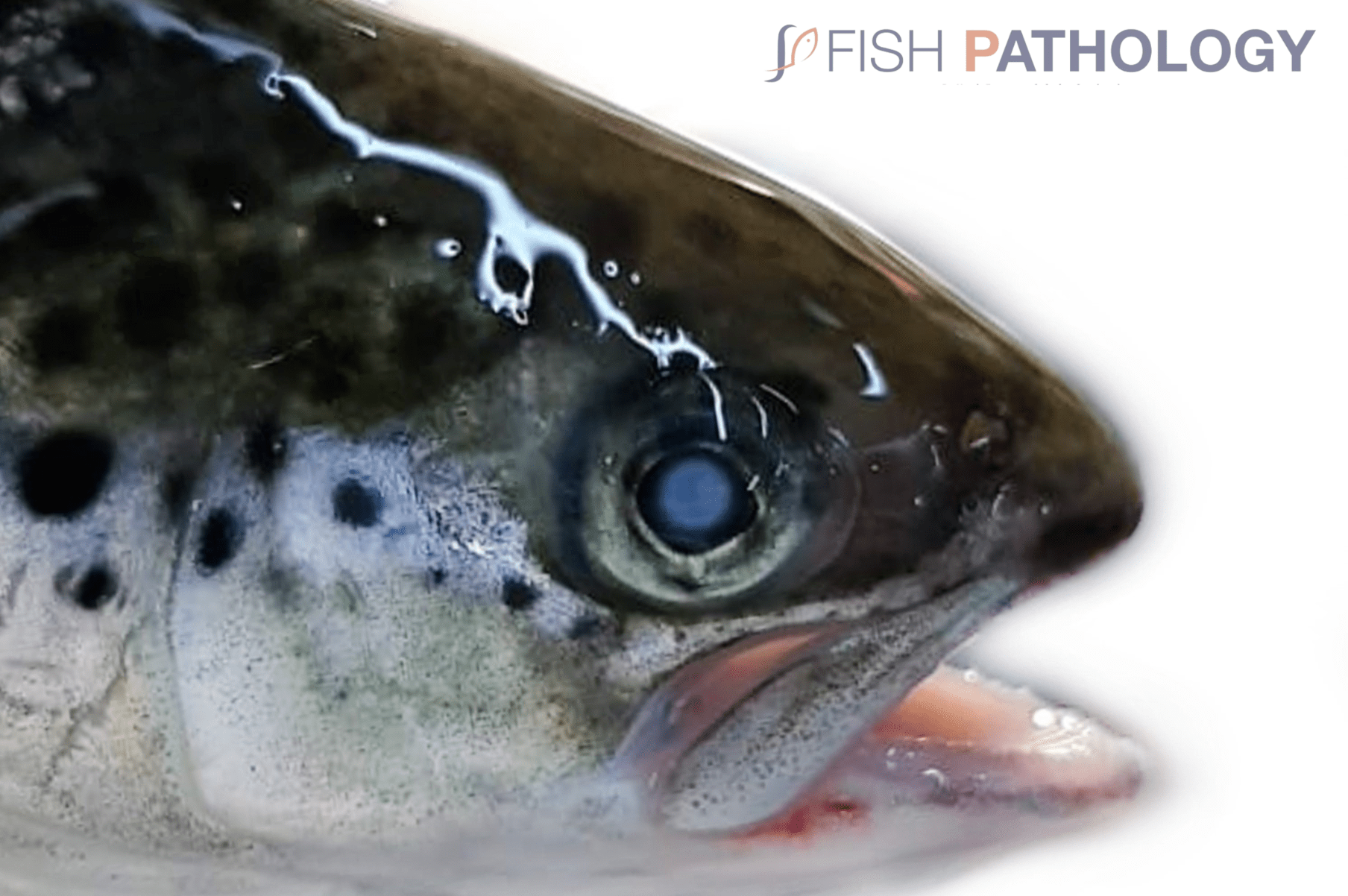
We have recently seen a high incidence of cataracts in farmed salmon in several European countries, Canada, and Chile. Salmonids depend on vision for normal feed intake and, not surprisingly, cataracts have been shown to reduce feed conversion efficiency and growth rates, which ultimately raise the cost of rearing fish. Consequently, cataracts can have a significant economic impact on salmonid aquaculture.
It is important to note at the outset that “cataracts” is a clinical definition, and their presence does not necessarily mean that changes can be seen histopathologically. Cataracts have a multifactorial aetiology including trauma, nutritional deficiency, ultra-violet radiation (UV or actinic damage), gas supersaturation, osmotic imbalances, environmental chemicals, adverse side-effects from drugs treatment, parasitic infestation, and toxins from intra-ocular bacterial infections.
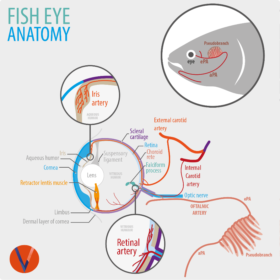
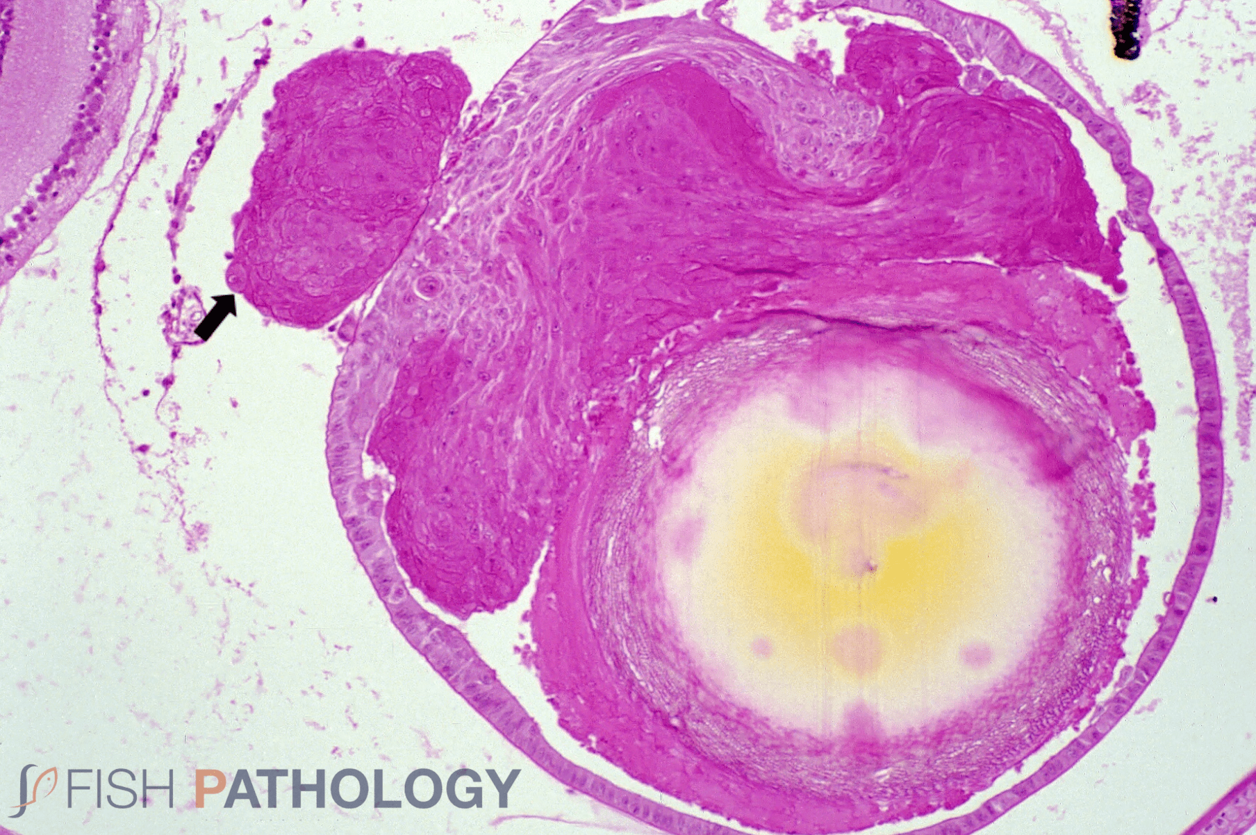
By contrast with mammals, the fish lens is spherical and unable to change shape. Accommodation (focusing) occurs, therefore, by moving the lens in and out of the light pathway by means of the retractor lentis muscle. The lens has a capsule with an underlying epithelium extending around it to varying degrees depending on the species. This epithelium is metabolically very active, one of its main functions being to desiccate the lens, thereby maintaining clarity. Anything that damages the epithelium or impairs this metabolic activity, therefore, leads to uptake of water, osmotic swelling, and loss of clarity of the lens (cataract).
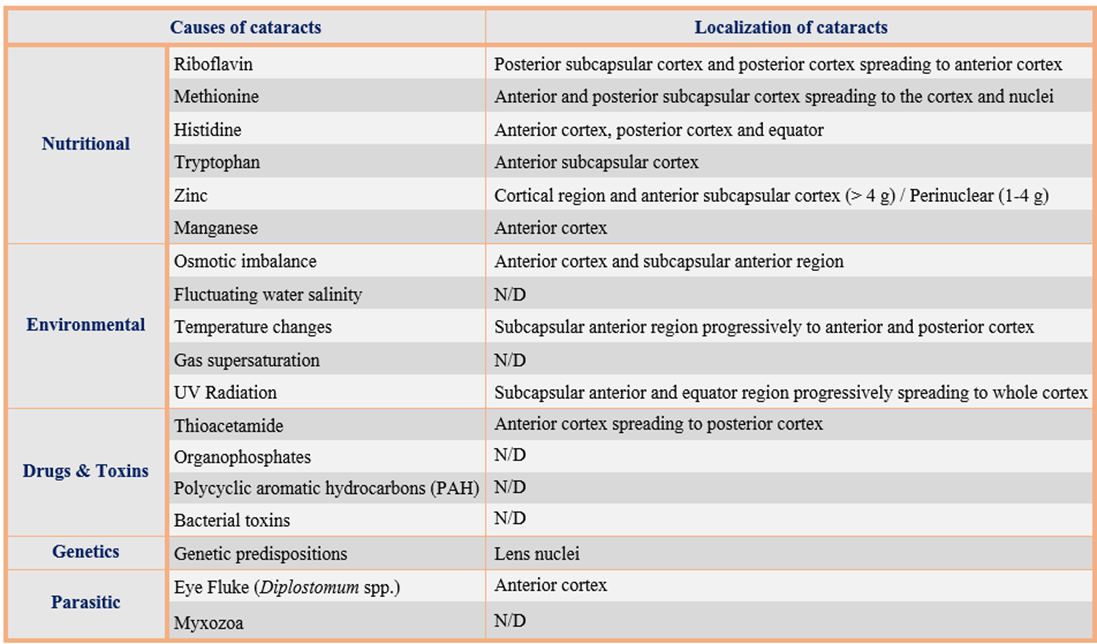
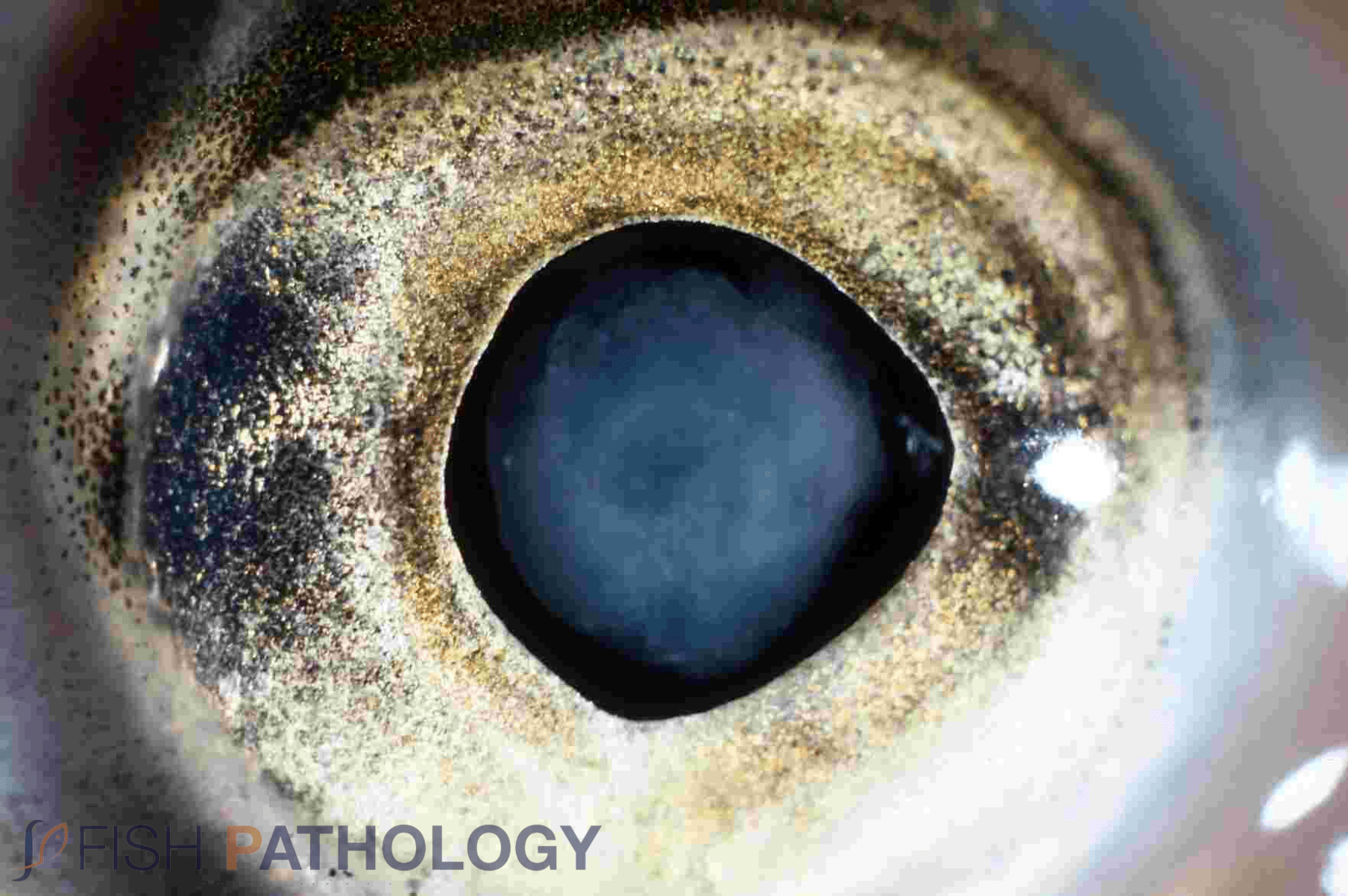
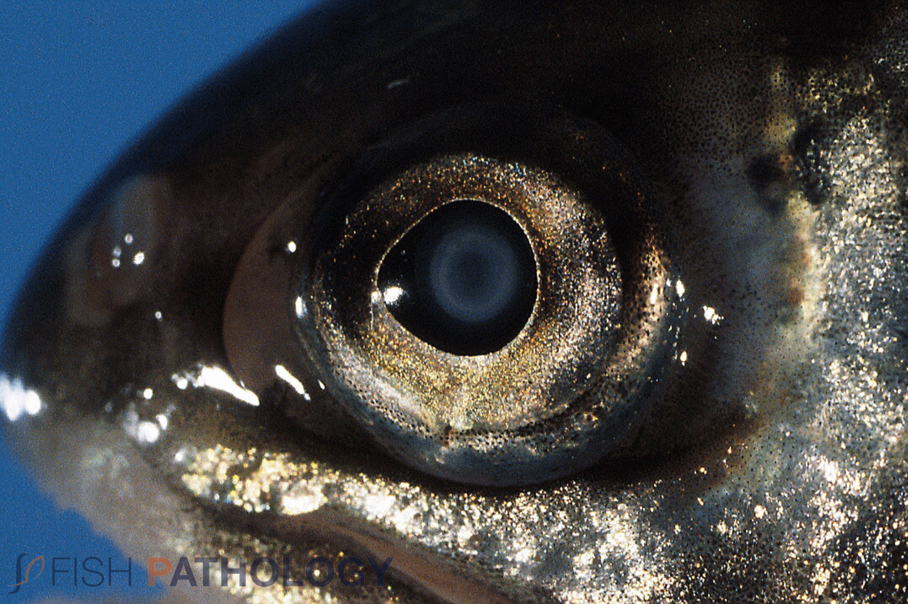
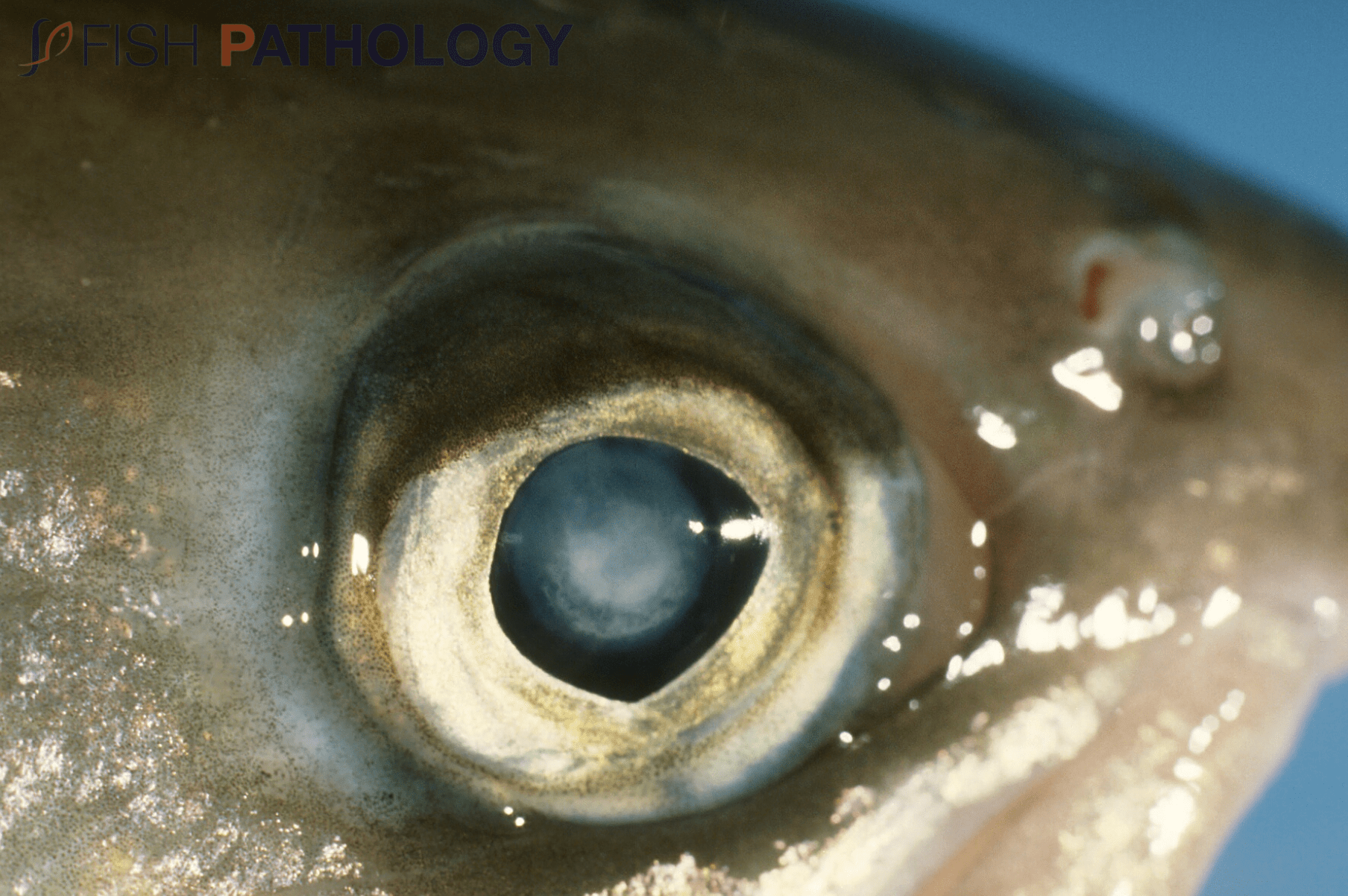
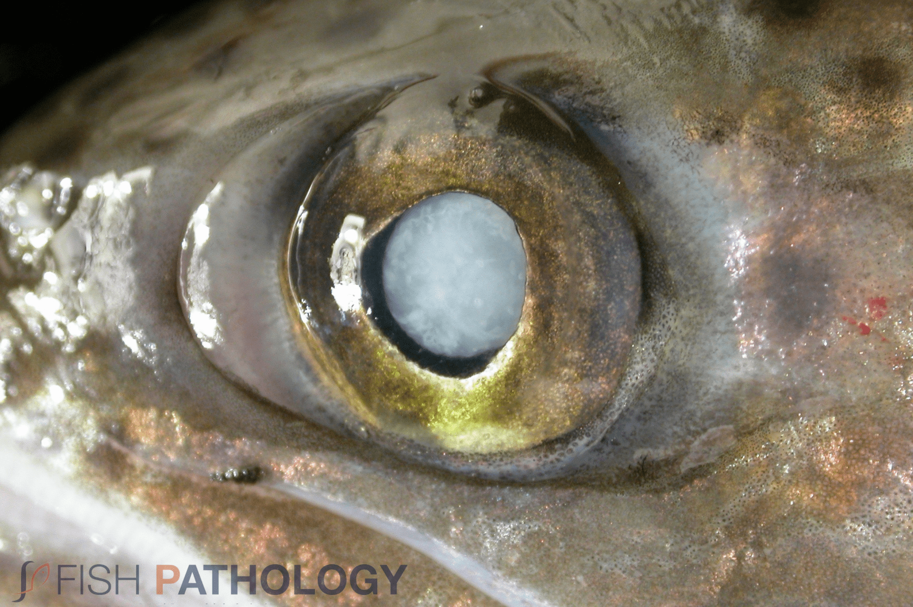
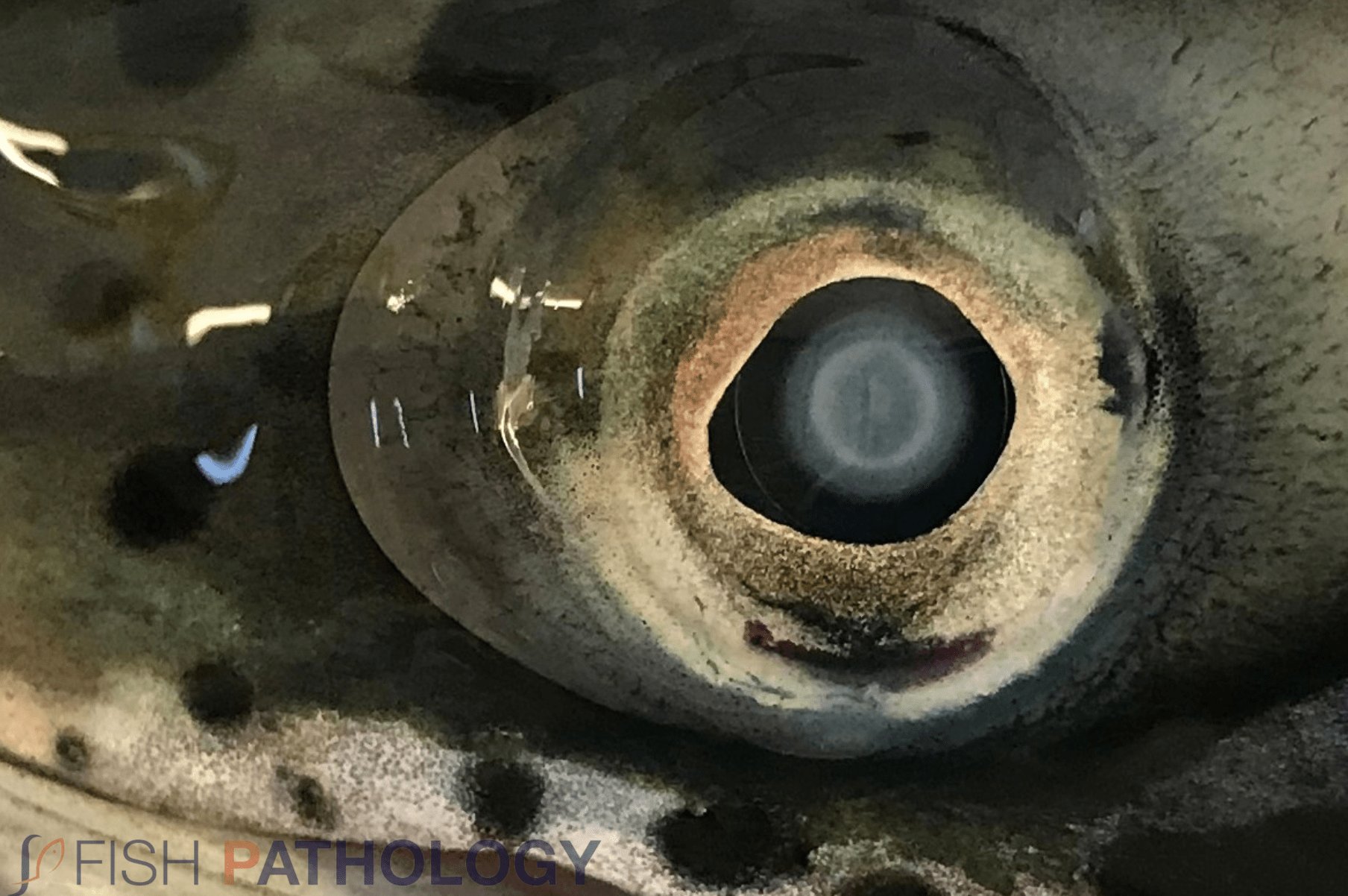
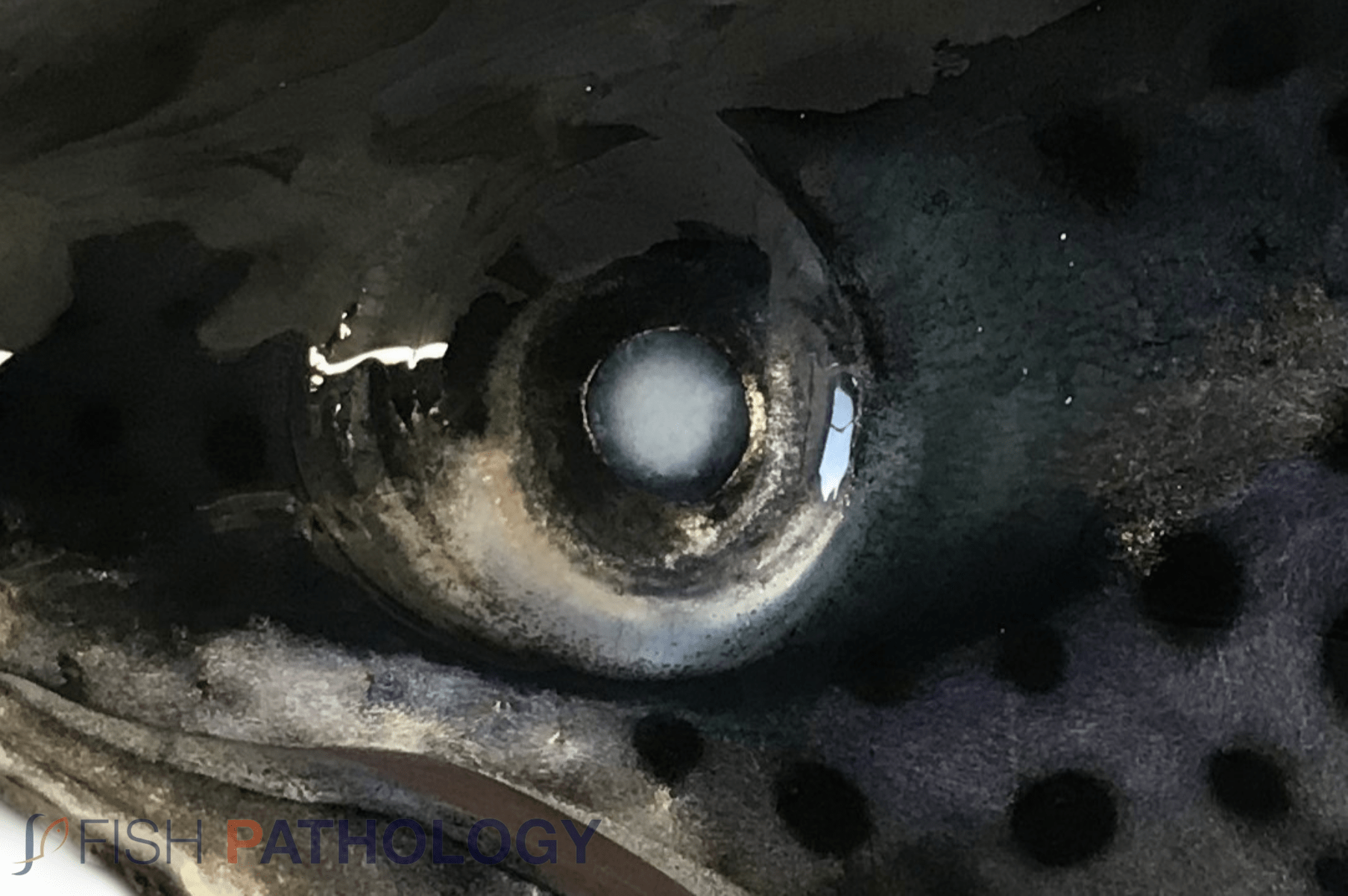
Cataracts are defined as opacities in the lens or the lens capsule that cause a reduced visual acuity. In early stages small white spots in the central part of the lens are observed and in more advanced stages the entire lens may be involved. Additionally, white “halos” in the lens can be observed. Cataracts can appear in different parts of the lens and the pattern of the changes can suggest an aetiology (see Table 1).
Depending on the aetiology, cataracts can be reversible or irreversible, although there are few definitive studies on this, and most authorities extrapolate from and use mammals for comparison. Given the remarkable regenerative powers of teleosts (including central nervous system and myocardium), such comparisons and extrapolations are dangerous! Nevertheless, and for example, osmotic cataracts in salmonids are known to be reversible if the damage is not too long-lasting or has caused disruption of the lens fibres. On the other hand, high experimental doses of UV radiation will produce irreversible cataract in trout.
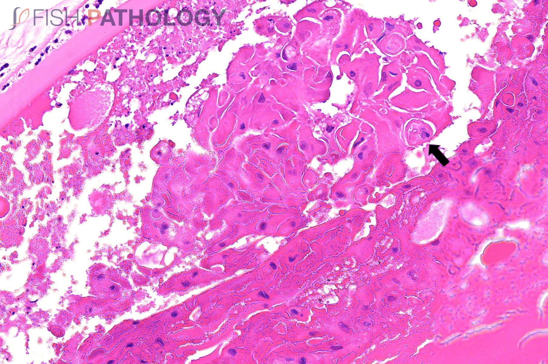
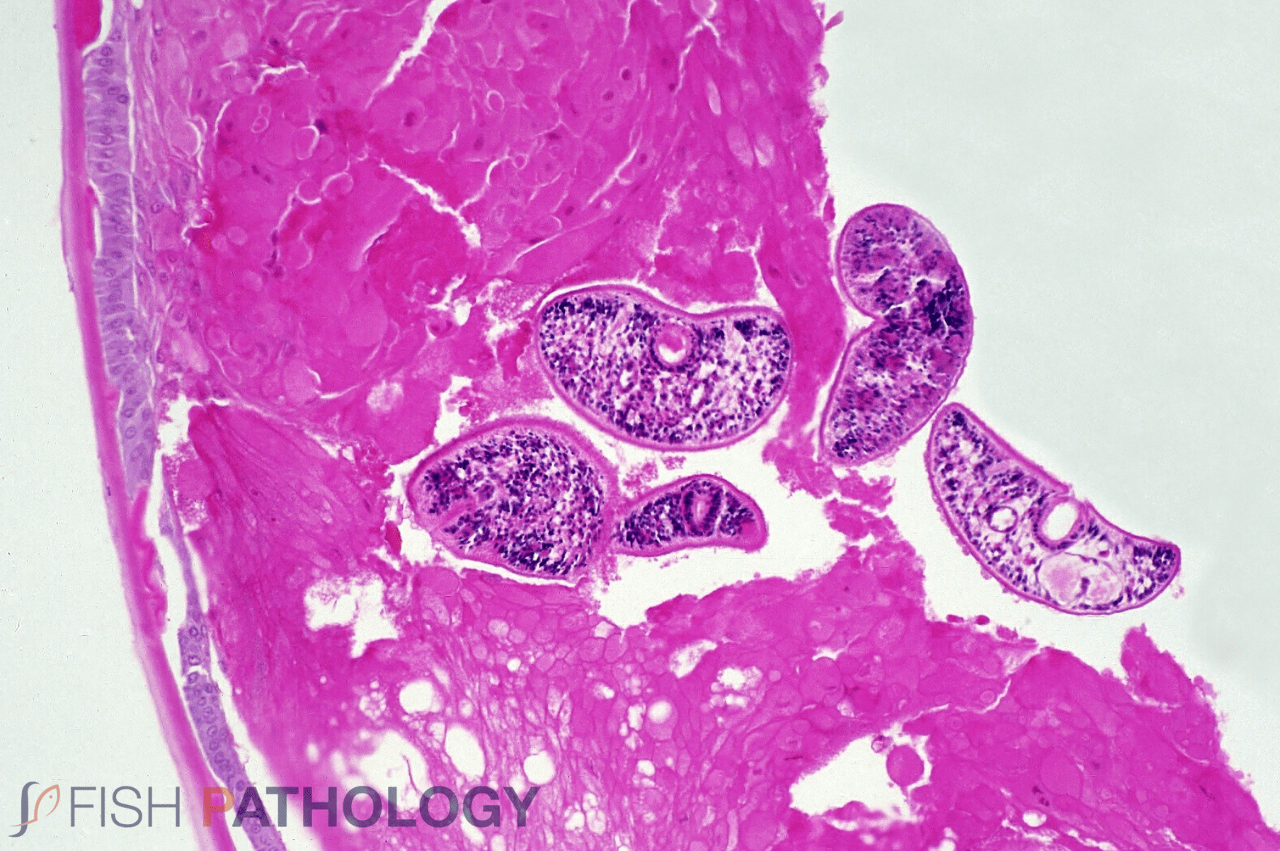
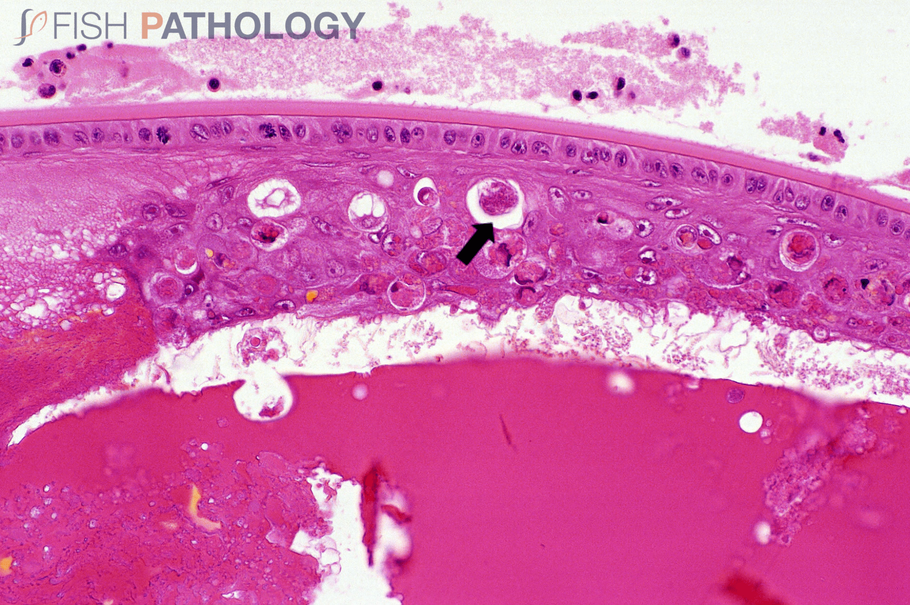
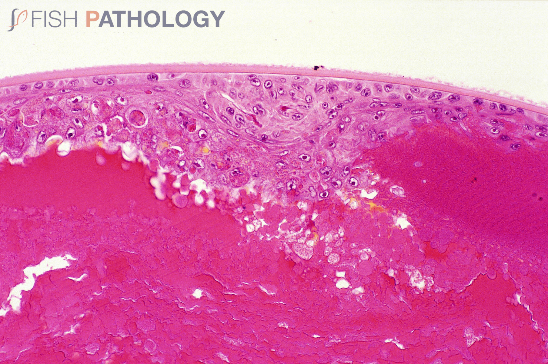
Histologically, opacities in the lens are characterized by lens fibres with abnormal features, including separation, swelling, granularity, condensation, fragmentation and liquefaction, all creating cortical “Morgagnian degeneration” (Morgagnian globules). Fibre necrosis and disruption of the normal configuration can also be seen as well as abnormal retention of fibre nuclei. Swollen lens fibres with abnormally retained nuclei are sometimes referred to as “bladder” or “balloon” cells.
Additional features associated with cataracts include thickening or mineralization of the lens capsule, vacuolation of subepithelial cortical fibres, epithelial cell proliferation and/or reduplication, and the presence of subepithelial lakes of proteinaceous material. The lens capsule can also rupture leading to release of lens material into the eye. In mammals this material induces severe inflammation; such is not invariably the case in fish.
IMAGES


REFERENCES
Bjerkås, E., Holst, J., & Bjerkås, I. (2004). Cataract in farmed and wild Atlantic salmon (Salmo salar L.). Animal Eye Research, 3-13.
Bjerkas, E., Holst, J., Bjerkas, I., & Ringvold, A. (2003). Osmotic cataracts causes reduced vision in wild Atlantic salmon postsmolts. Diseases of Aquatic Organisms, 151-159.
Bjerkås, E., Waagbø, R., Sveier, H., Breck, O., Bjerkås, I., Bjørnestad, E., & Maage, A. (1996). Cataract Development in Atlantic salmon (Salmo salal L.) in Fresh Water. Acta Veterinaria Scandinavica, 351-360.
Doughty, M., Cullen , A., & Monteich-McMaster, C. (1997). Aqueous humour and crytalline lens changes associated with ultraviolet radiation or mechanical damage to corneal epithelium in freshwater rainbow trout eyes. Journal of Photochemistry and Photobiology , 165 – 172.
Ersdal, C., Midtlyng, P., & Jarp, J. (2001). An epidemiological study of cataracts in seawater farmed Atlantic salmon Salmo salar. Disease of Aquatic Organisms, 229-236.
Ersdal, C., Midtlyng, P., & Jarp, J. (2001). An epidemiological study of cataracts in seawater farmed Atlantic salmon Salmo salar. Diseases of Aquatic Organisms, 229-236.
Ferguson, H. (2006). Systemic Pathology of Fish: a text and atlas of normal tissues in teleosts and their responses in disease. London: Second Edition. Scotian Press.
Hargis, W. J. (1991). Disorders of the eye in finfish. Fish Diseases , 95-117.
Midtlyng, P. J., Ahrend, M., Bjerkås, E., Waagbø, R., & Wall, T. (1999). Current research on cataracts in fish. Bulletin of the European Association of Fish Pathologists, 299.
Peachey, B. L., Scott, E. M., & Gatlin, D. M. (2017). Dietary histidine requirement and physiological effects of dietary histidine deficiency in juvenile red drum Sciaenop ocellatus. Aquaculture, 244-251.
Remø, S. C., Hevrøy, E. M., Breck, O., Olsvik, P. A., & Waagbø, R. (2017). Lens metabolic profiling as a tool to understand cataractogenesis in Atlantic salmon and rainbow trout reared at optimum and high temperature. Journal Plos One , 1-21.
Rhodes, J., Breck, O., Waagbø, R., Bjerkås, E., & Sanderson, J. (2010). N-acetylhistidine, a novel osmolyte in the lens of Atlantic salmon (Salmo salar L.). American Journal of Physiology, 1075-1081.
Sambraus, F., Fjelldal, P., Remø, S., Hevrøy, E., Nilsen, T., Thorsen, A., Waagbø, R. (2017). Water temperature and dietary histidine affect catarct formation in Atlantic salmon (Salmo salar L.) diploid and triploid yearling smolt. Journal of Fish Diseases, 1195-1212.
Waagbø, R., Bjerkås, E., Sveier, H., Breck, O., Bjørnstad, E., & Maage, A. (1996). Nutritional status assessed in groups of smolting Atlantic salmon, Salmo salar L., developing cataracts. Journal of Fish Diseases, 365-373.
Waagbø, R., Tröße, C., Koppe, W., Fontanillas, R., & Breck, O. (2010). Dietary histidine supplementation prevents cataract development in adult Atlantic salmon, Salmo salar L., in seawater. British Journal of Nutrition , 1460-1470.
Wall, A. E. (1998). Cataracts in farmed Atlantic salmon (Salmo salar) in Ireland, Norway and Scotland from 1995 to 1997. The Veterinary Record, 626-631.
