Rainbow trout, multi-focal granulomatous dermatitis

Spawning Rash – Gross Pathology

The bacterium Renibacterium salmoninarum is a small (~ 1.0 μm), intracellular, non-motile diplobacillus, gram +, that is slow growing and a fastidious pathogen.

Rainbow trout, multi-focal granulomatous dermatitis
Figure 1. Rainbow trout, multi-focal granulomatous dermatitis.

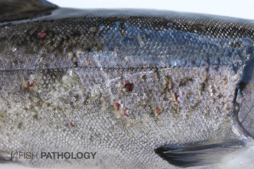
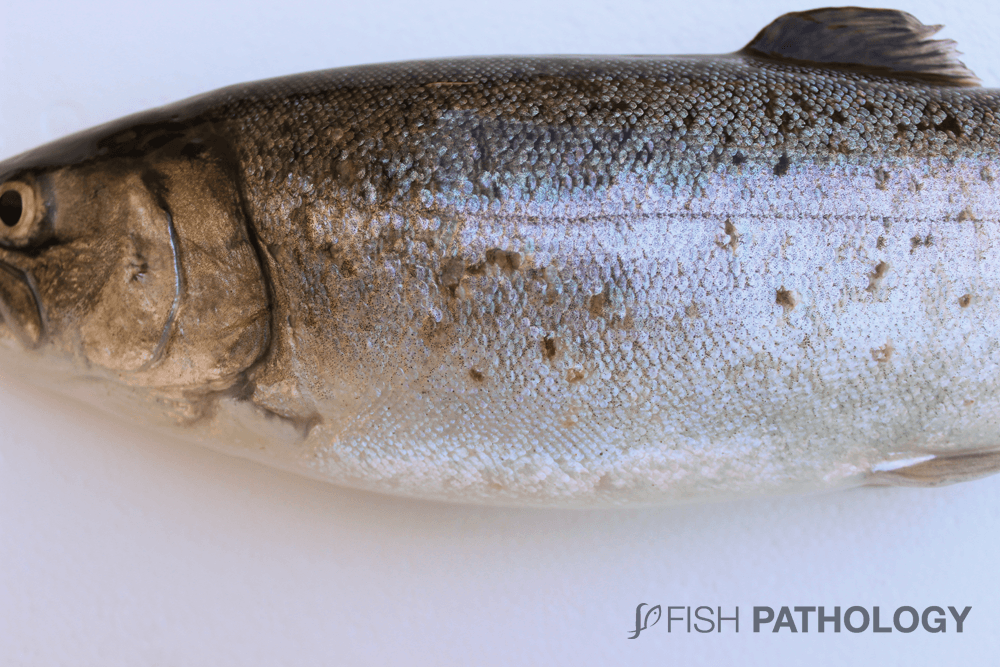
The external gross findings on the skin could be a good example of chronic-active dermal o subdermal lesion associated with R. salmoninarum.


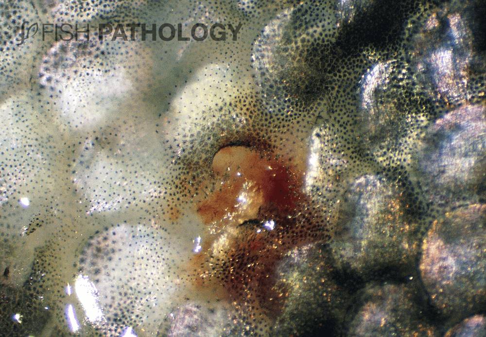
“Spawning rash” can be observed in adults, particularly at or around spawning time. Lesions may be largely dermal, often centering round scale-pockets to produce multifocal chronic-active dermatitis. Alternatively the response, mainly in the superficial muscle, may be so severe as to dissect the whole dermis away from the subcutis to produce large fluid-filled cavitations or blisters.

O. kisutch. Reversal of scales and formation of multifocal ulcers are observed, consistent with dermal infection by R. salmoninarum.
Figure 2. Coho salmon. Reversal of scales and formation of multifocal ulcers are observed, consistent with dermal infection by R. salmoninarum.


Dermal lesions may occur in the absence of detectable lesions elsewhere in the body, including the kidney, and they resolve after spawning. This time-limited susceptibility illustrates the immunological changes seen in the skin at spawning.


The cellular response involves mainly neutrophils and macrophages and is a good example of chronic-active dermatitis.

Rainbow trout, multi-focal granulomatous dermatitis
Figure 3. Rainbow trout, multi-focal granulomatous dermatitis.
O. kisutch. Reversal of scales and formation of multifocal ulcers are observed, consistent with dermal infection by R. salmoninarum.
Figure 4. Coho salmon. Reversal of scales and formation of multifocal ulcers are observed, consistent with dermal infection by R. salmoninarum.


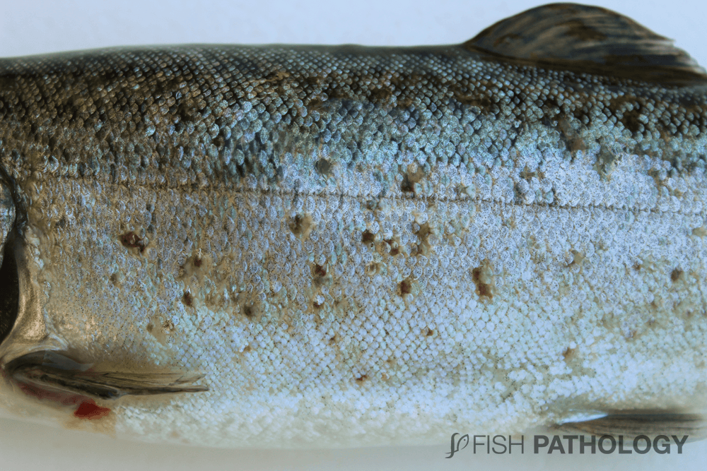
At gross pathology, in the initial phases of this disease, small elevations of the scales are observed with subsequent inversion in their position and detachment. Subsequently, ulceration and haemorrhage of the integument occur.

O. kisutch. Reversal of scales and formation of multifocal ulcers are observed, consistent with dermal infection by R. salmoninarum.
Figure 5. Coho salmon. Reversal of scales and formation of multifocal ulcers are observed, consistent with dermal infection by R. salmoninarum.


REFERENCES

  • Benediktsdóttir, E., Helgason, S., & Gudmundsdóttir, S. (1991). Incubation time for the cultivation of Renibacterium salmoninarum from Atlantic salmon, Salmo salar L., broodfish. Journal of fish diseases14(1), 97-102.
  • Ferguson, H. W., 2006, Systemic Pathology of Fish, London, UK, Scottian Press.

By: Hugh Ferguson

Dr Ferguson earned his veterinary degree from the Royal (Dick) School of Veterinary Studies, Edinburgh, Scotland and held a Wellcome Research Fellowship at the Institute of Aquaculture, Stirling University where he obtained his PhD. He then worked for 4 years as a diagnostic pathologist at the Veterinary Research Laboratories, Belfast, Northern Ireland, prior to moving to Canada. He left Ontario Veterinary College after 19 years as a full professor of veterinary pathology, to return to Scotland to become head of diagnostic pathology in Stirling. During all this time he became board-certified in the American college of veterinary pathology (ACVP), and a Fellow of the Royal College of Pathologists (FRCPath, London). After Scotland he moved to become chair of veterinary pathology, and Senior Research Fellow in Windward Islands Research and Education Foundation (WINDREF), St George’s University (SGU), Grenada, West Indies. He has published more than 230 papers in refereed journals.

totop