Atlantic salmon (adult-1500 g) with BKD. Note the multiple granulomas in kidney. These severe lesions lead to renal failure

Bacterial Kidney Disease (BKD) in Salmonids – Gross Pathology

Bacterial kidney disease (BKD) is a chronic systemic infection of salmonid fish, the cause of which is the gram-positive diplobacillus, Renibacterium salmoninarum. Although BKD is most common in salmonids, both farmed and wild, R. salmoninarum is also found in other fish species, such as cyprinids and sablefish Anoplopoma fimbria.

R. salmoninarum grows intracellularly in the phagocytic cells of the fish, resulting therefore in a largely granulomatous host response, although the early response to infection does involve significant numbers of neutrophils.

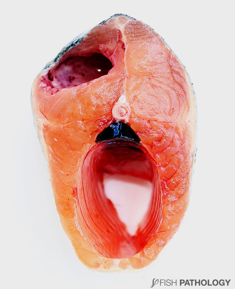
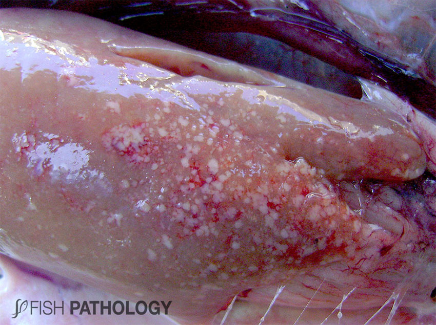
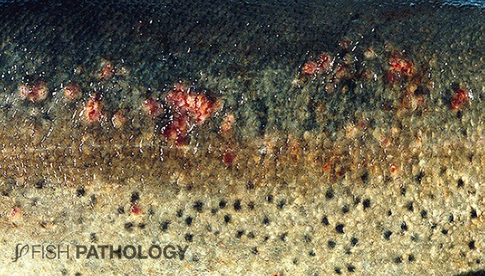
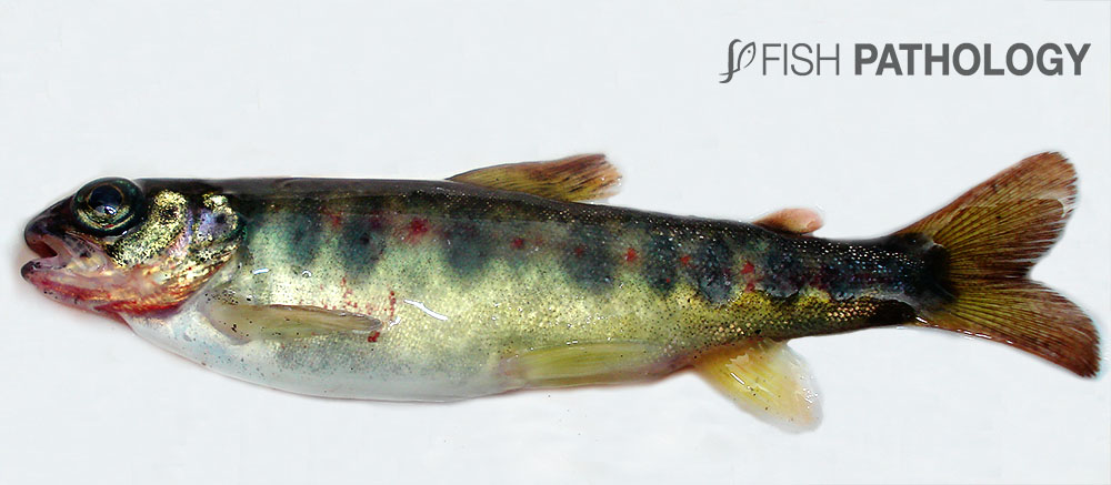
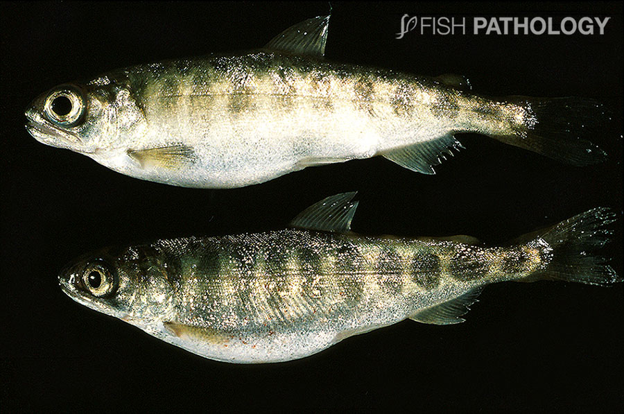
Clinical signs and external lesions are variable and range from darkening and a mottled appearance to the skin, distended abdomen, exophthalmia, petechiae and haemorrhaging around the base of the pectoral fins and the lateral line. Superficial blister with vesicle formation, abscessation and subsequent ulceration may develop in the skin and even cavitations in the musculature can form. Nervous symptoms are encountered if the brain becomes involved; this is not uncommon if the fish are fed long-term antibiotics (those which cannot cross the blood-brain barrier).

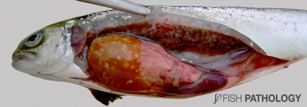
Gills and internal organs can be pale, indicating the anaemia that is often present. Given the extent of involvement and destruction of haemopoietic tissue in kidney and spleen, this is probably a myelophthisic anaemia.

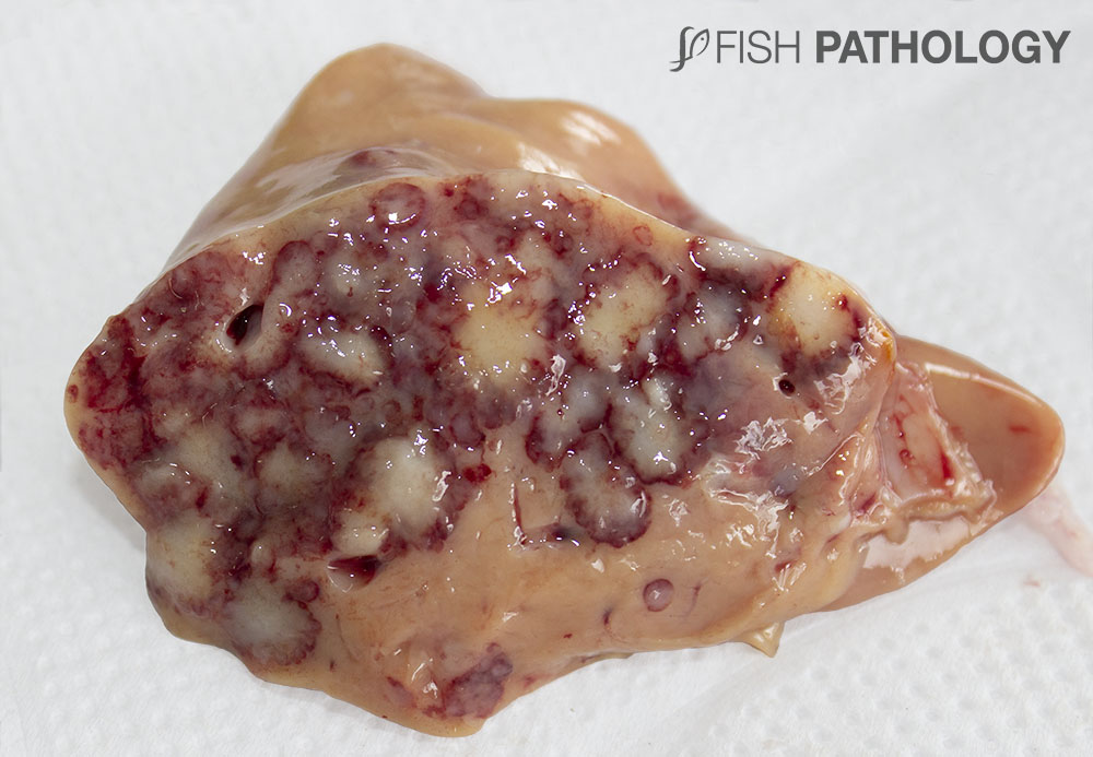
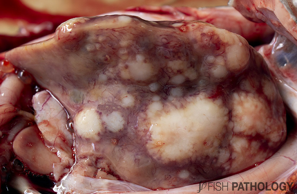
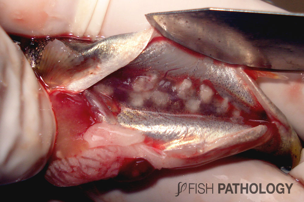
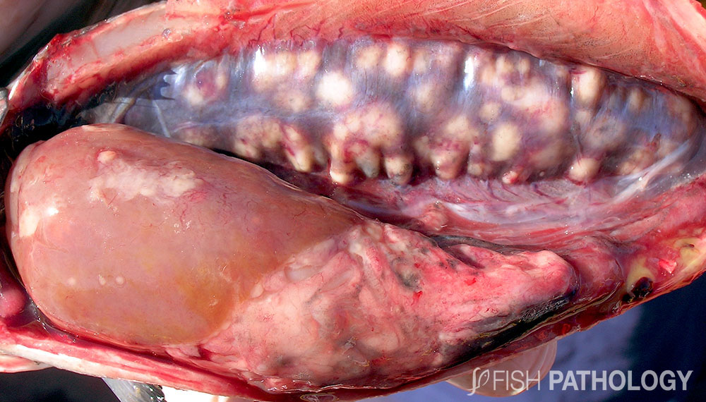
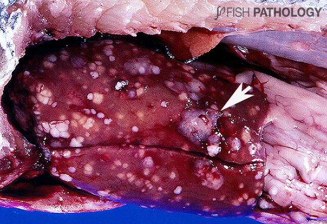
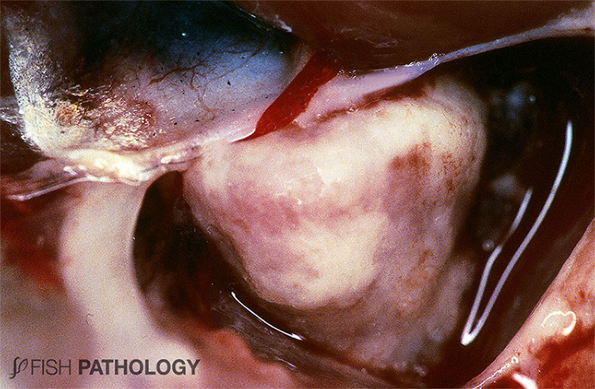
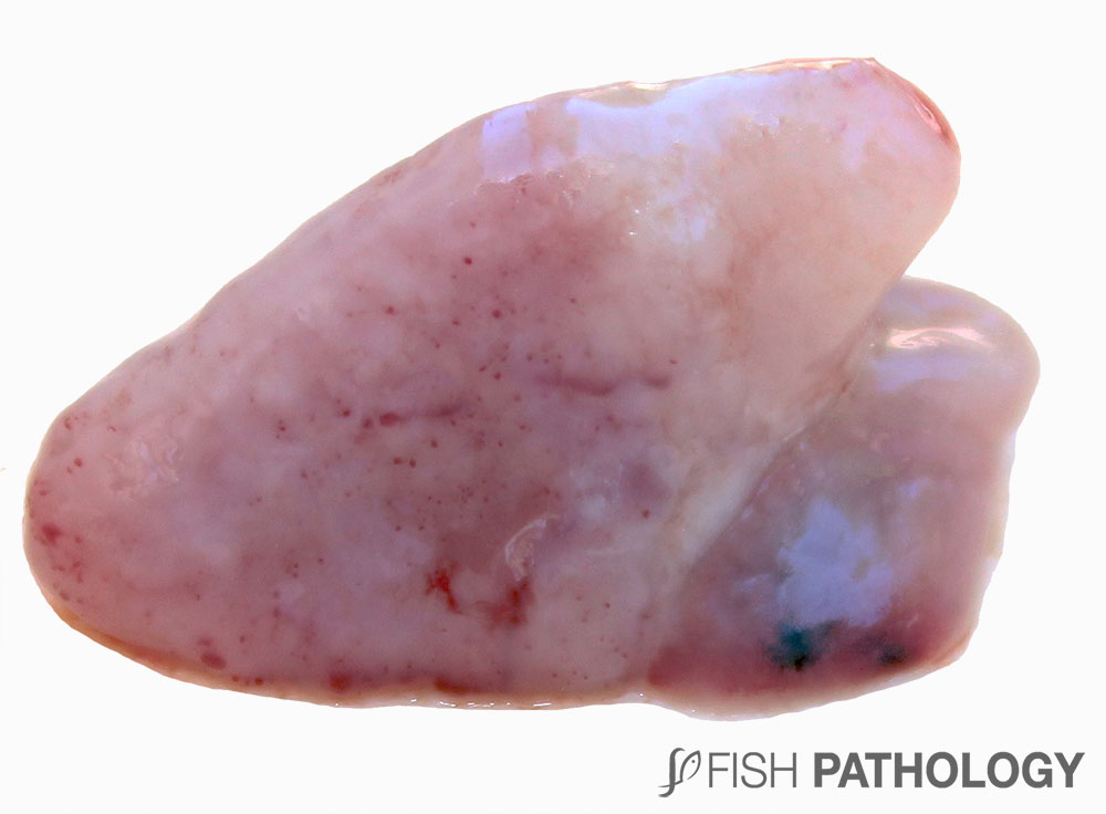
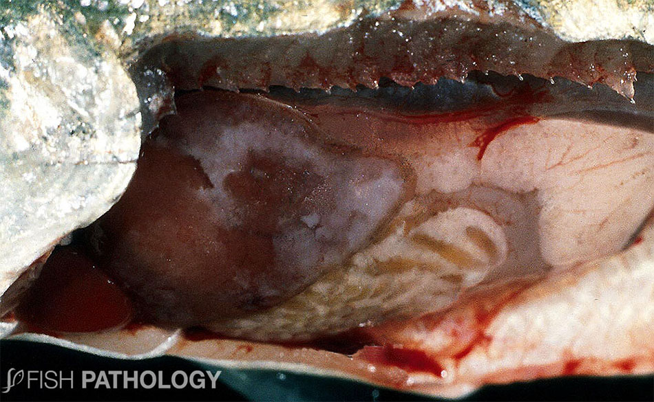
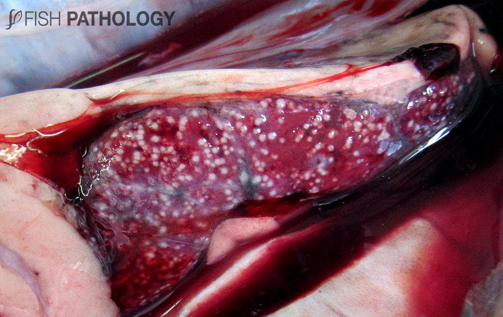
Granulomas are systemic and can be observed in spleen, heart, liver, and of course the kidney from which the disease derives its name. The kidney is generally swollen, and grey-white granulomas of various sizes are seen on the surface of and throughout the organ. Perforation of the overlying renal capsule results in peritonitis, with petechial haemorrhaging of peritoneum and underlying muscle. Peritonitis and/or destruction of sufficient nephrons (impairing fluid balance) can result in an accumulation of peritoneal fluid and a distended abdomen. Peritonitis can lead to the formation of diphtheritic membranes covering all internal organs, most easily seen over the liver and spleen. The heart can be so severely involved as to be almost obliterated, with pericarditis leading to constriction and cardiac tamponade as common sequelae. A yellow, viscous fluid occurs in the intestine and may contain blood.

A seasonal “spawning rash” has been reported in adults around spawning time, where a pustular dermatitis may cover large areas of the skin with many blisters or raised haemorrhagic nodules within the epidermis. Granulomatous tissue invades adjacent scale pockets and extends longitudinally along the fibrous tissue layer of the dermis. These lesions disappear after spawning.

Figure 1. Rainbow trout with BKD. Note the multiple granulomas in liver.
Figure 2. Rainbow trout with BKD. This is the same fish as shown in Figure 1. Note the multiple granulomas in liver.
Figure 3. Coho salmon with BKD. Note cavitation in the musculature.
Figure 4. Atlantic salmon (pre-smolt) with BKD. Note the multiple granulomas in liver and kidney (Picture courtesy of Jaime Santana).
Figure 5. Salmonid with BKD. Note the haemorrhage and multiple granulomas in liver.
Figure 6. Atlantic salmon fingerling with BKD. Note the multiple granulomas in kidney. Such severe lesions lead to renal failure with impaired fluid balance.
Figure 7. Atlantic salmon (adult-1500 g) with BKD. Note the multiple granulomas in kidney. These severe lesions lead to renal failure (Picture courtesy of Marcelo Vera).
Figure 8. Atlantic salmon with BKD. Note the presence of white nodules (granulomas) on the gill (Picture courtesy of Iván Ñancufil).
Figure 9. Rainbow trout with multifocal granulomatous hepatitis due to BKD. Lesions are present on and just beneath the capsular surface, as well as throughout the parenchyma. One or two granulomas had formed adhesions to the peritoneal lining and these were “torn” when the fish was opened up at necropsy, leaving tags on the capsular surface (arrow).
Figure 10. Severe chronic fibrinous pericarditis in Pacific salmon due to BKD. Such severe constrictive lesions lead to impaired cardiac output (cardiac tamponade).
Figura 11. Pericarditis fibrinosa temprana en salmón Coho debido a BKD.
Figure 11. Early fibrinous pericarditis in Coho salmon due to BKD.
Figure 12. Fibrinous peritonitis in salmon with BKD leading to a white diphtheritic membrane on the surface of the liver.
Figure 13. Rainbow trout with BKD showing multi-focal granulomatous dermatitis. This lesion is often referred to as “spawning rash” – it disappears after spawning.
Figure 14. Atlantic salmon (fingerling) with BKD. Note the exophthalmia (a consequence of loss of osmotic integrity due to severe renal destruction) and the sub-opercular and mandibular haemorrhage.
Figure 15. Coho salmon fingerlings with BKD showing exophthalmia and peritoneal distension.
Figure 16. Atlantic salmon with BKD. Note the multiple granulomas in spleen (Picture courtesy of Antonia Sabugal).
totop