
Flavobacterium psychrophilum is a ubiquitous Gram-negative filamentous and yellow-pigmented bacterium, and as the name suggests, it thrives in cooler water temperatures, causing disease in freshwater fish at 4-12°C.
It likes connective tissues, especially those in younger fish in which it causes diseases such as bacterial cold-water disease (BCWD or peduncle disease) and rainbow trout fry syndrome (RTFS).
As fish get older and the proportions/chemistry/locations of connective tissue change, the types of lesions also change.
F. psychrophilum is responsible for significant economic losses in salmonid aquaculture in freshwater. Coho salmon and rainbow trout appear to be particularly susceptible and losses can be extremely high.
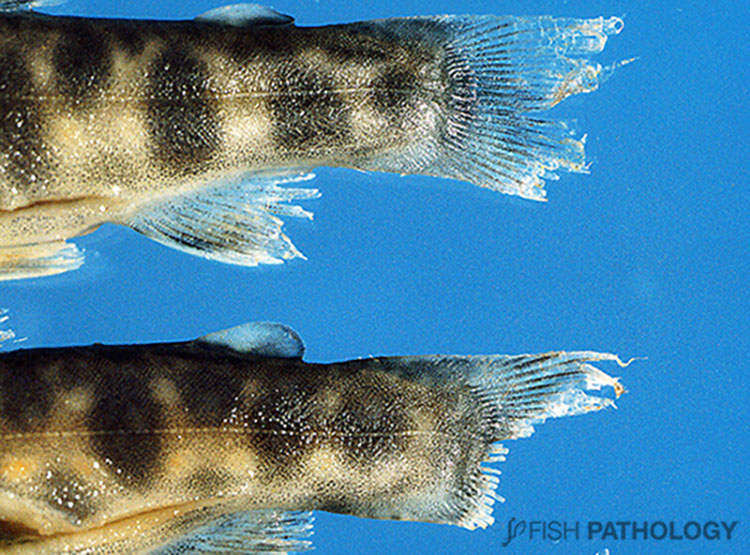
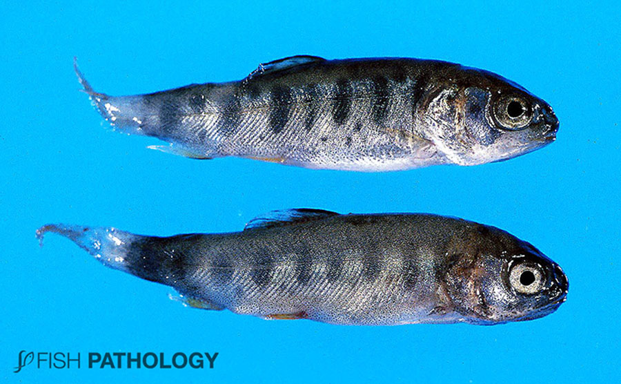
BCWD is a largely superficial disease involving fins and dermis. The first changes in a population are usually seen in the caudal fin which develops a white rim to its margin.
This can be followed by progressive necrosis and involvement of the whole peduncle which can darken (peduncle disease).

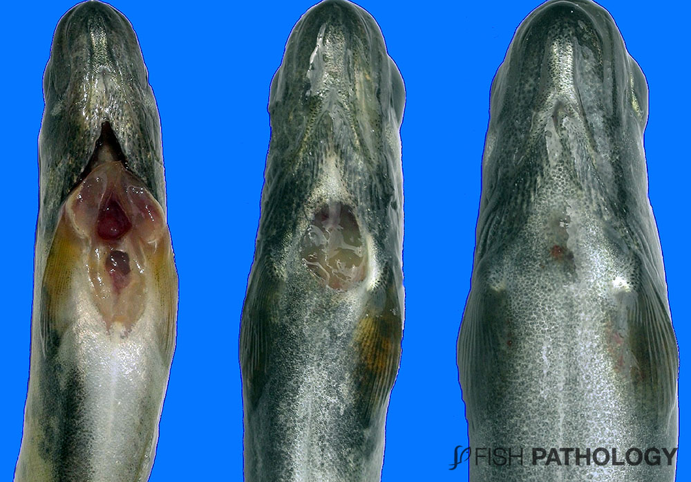
In other situations, infection with F. psychrophilum can be more systemic (e.g. RTFS), with involvement of cartilage throughout the body, including that surrounding the brain, eyes, gills and spinal column.
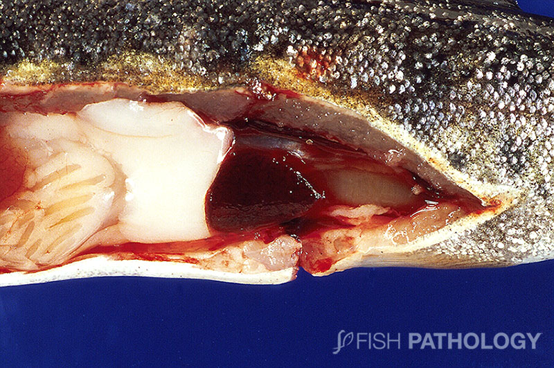
As may be imagined from this tissue predilection, gross lesions may include panophthalmitis, pallor of the gills and spinal deformity. Splenic enlargement may also be seen, often with a degree of peritonitis and ascites, leading to abdominal distension.
If cranial cartilages are severely involved, or those of the spinal column, fish may exhibit nervous dysfunction, leading to concerns about other diseases that may target the cranial cartilage such as whirling disease. If gill cartilage is severely affected, fish can sometimes show respiratory distress.
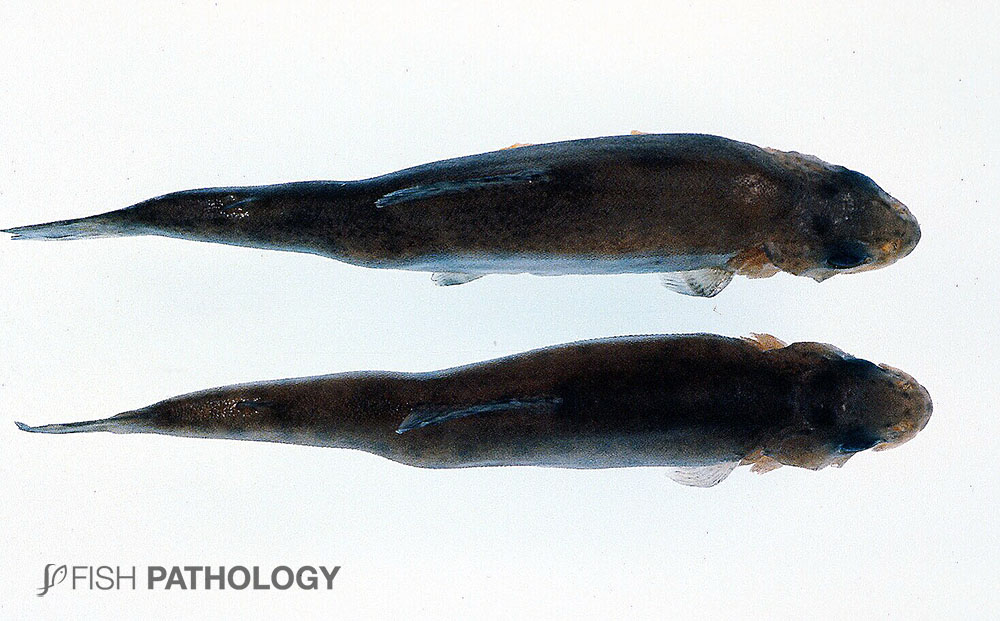
In Coho salmon, which are highly susceptible, the skin covering the yolk sac of fry may be eroded and mortalities as high as 50% may occur.
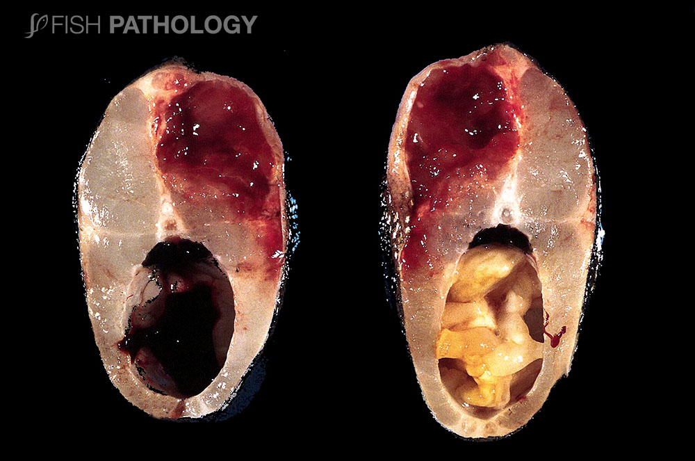
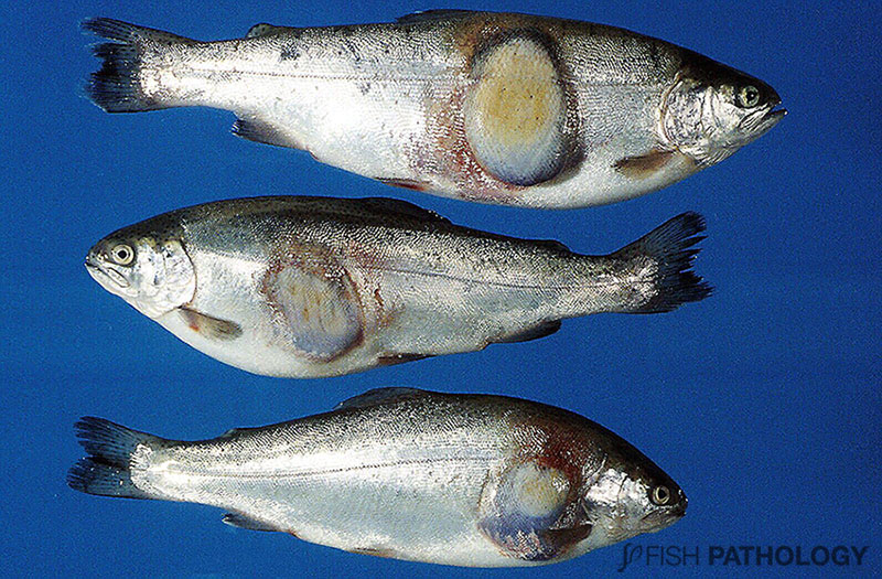
In older fish, muscle can sometimes be targeted, leading usually to focal but severe haemorrhagic and necrotic myositis, again a differential for other bacterial diseases such as furunculosis.
F. psychrophilum has also been causally linked to the severe but non-fatal skin condition of rainbow trout “Strawberry disease”, but so too have other bacteria.
REFERENCES
- Barnes, M. E., & Brown, M. L. (2011). A review of Flavobacterium psychrophilum biology, clinical signs, and bacterial cold water disease prevention and treatment. Open Fish Science Journal, 4, 40.
- Ekman, E., & Norrgren, L. (2003). Pathology and immunohistochemistry in three species of salmonids after experimental infection with Flavobacterium psychrophilum. Journal of fish diseases, 26(9), 529-538.
- Ferguson, H.W., 2006, Systemic Pathology of Fish, London, UK, Scotian Press.
- Jarau, M., Di Natale, A., Huber, P. E., MacInnes, J. I., & Lumsden, J. S. (2018). Virulence of Flavobacterium psychrophilum isolates in rainbow trout Oncorhynchus mykiss (Walbaum). Journal of fish diseases, 41(10), 1505-1514.
- Madsen, L., Møller, J. D., & Dalsgaard, I. (2005). Flavobacterium psychrophilum in rainbow trout, Oncorhynchus mykiss (Walbaum), hatcheries: studies on broodstock, eggs, fry and environment. Journal of fish diseases, 28(1), 39-47.
- Nematollahi, A., Decostere, A., Pasmans, F., & Haesebrouck, F. (2003). Flavobacterium psychrophilum infections in salmonid fish. Journal of fish diseases, 26(10), 563-574.
- Nilsen, H., Johansen, R., Colquhoun, D. J., Kaada, I., Bottolfsen, K., Vågnes, Ø., & Olsen, A. B. (2011). Flavobacterium psychrophilum associated with septicaemia and necrotic myositis in Atlantic salmon Salmo salar: a case report. Diseases of aquatic organisms, 97(1), 37-46.
- Noga, E.J., 2000, Fish Disease – Diagnosis and Treatment, Iowa, USA, Blackwell Publishing company.
- Roberts, R.J., 2012, Fish Pathology, Oxford, USA, Blackwell Publishing Ltd.
