
Gas bubble disease (GBD) is the fish equivalent of the “bends” in human scuba divers. It is the result of gas coming out of solution in the bloodstream, thereby leading to the formation of emboli, especially in smaller blood vessels, including rete. Thus, lesions are common in gills, and the choroid gland of the eye. In water that is supersaturated, fish equilibriate with that supersaturated gas, just as human scuba divers equilibriate with gas at high pressures. It is not known precisely what prompts the gas to come out of solution in the bloodstream, but the drop in pressure seen as blood passes through small vessels is one likely possibility. As well as impeding blood flow, the gas bubbles can cause pressure necrosis of the vascular endothelium, and subsequent thrombosis. This is commonly seen in the gills.
Situations leading to supersaturation, and therefore where GBD is possible, include those where water warms up rapidly, and/or there is a sudden reduction in pressure. Both of these parameters conspire to reduce the solubility of gas in water. The analogy is sometimes drawn of a bottle of champagne being warmed up and then having its cork popped! Gas bubbles quickly appear! Disturbing/shaking the water (or champagne) can help to drive off the supersaturated gas.

In aquaria and hatcheries, GBD may be caused by leaks in pump manifolds or valve systems, air being “sucked in” and forced into solution – the so-called Venturi principle. Alternatively, it can be seen in fish being transported by air, a consequence of altitude (and therefore pressure) changes. In the wild, large algal blooms generating high levels of oxygen by photosynthesis have also been blamed, especially if the water remains calm.
Fish may die from GBD without any overt clinical signs, and diagnosis can be difficult; despite a detailed history, supersaturation can be a fleeting event, and not present when samples are collected. Sometimes “candling” fish in a strong light helps to spot gas bubbles, but these can be hard to see, even histopathologically. Mortality rate varies with the age and species of fish involved.

Sac fry with GBD are often forced to the water surface because of increased buoyancy. Gas bubbles can form between the yolk and the perivitelline membrane, as well as in the abdominal cavity, fins and cranium. Depending on the location of the bubble(s), affected fry could be head-up, tail-up or belly-up at the water surface.
In juveniles and adults, it is common for fish with acute GBD to die without showing visible lesions. Clinical behavioural signs in acutely affected animals include a sharp reduction in feeding, lethargy, loss of equilibrium and of buoyancy, aimless swimming, whirling with interspersed periods of inactivity, and spasmodic convulsions.
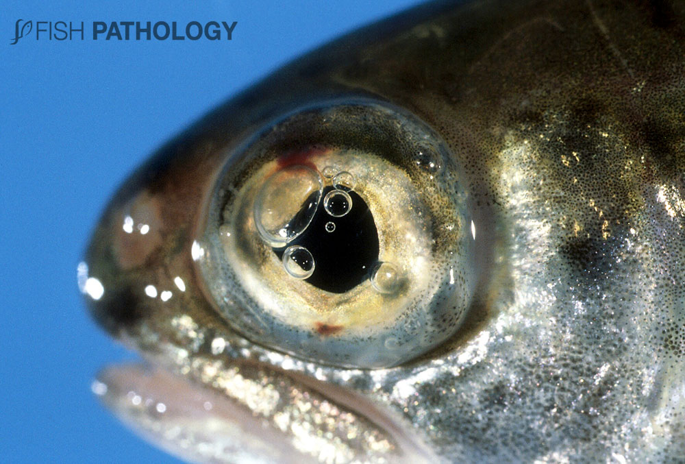
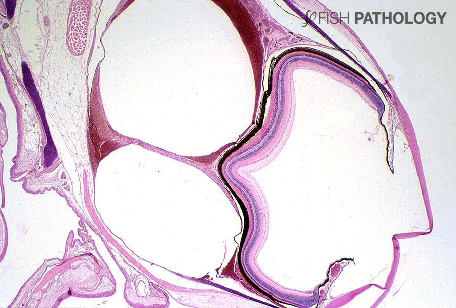
Uni- or bilateral exophthalmos is a classical sign of chronic GBD in juvenile and adult fish but is not invariably present. When exophthalmos exists, bubbles can be seen in all chambers of the eye and the sclera. The end result of the damage caused by expansion of the gas bubbles within the eye is often blindness and ultimately, phthisis.
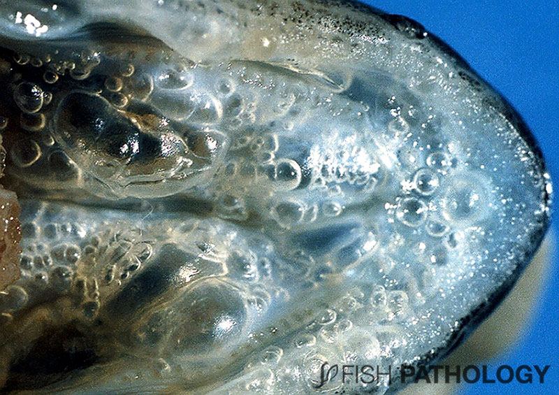
An interesting feature frequently noted in chinook salmon with GBD is the presence of gas bubbles within the blood vessels and soft tissues of the oral cavity – usually the roof of the mouth. These can cause fish to “cough” and vigorously work their opercula.
REFERENCES
- Bouck, G. R. (1980). Etiology of gas bubble disease. Transactions of the American Fisheries Society, 109(6), 703-707.
- Ferguson, Hugh W. (2006). Systemic Pathology of Fish, London, UK, Scotian Press.
- Leatherland, John F., Patrick Woo T.K (2010). Fish Diseases and Disorders, Volume 2: Non-infectious Disorders. Oxfordshire, UK, CABI.
- Machova, J., Faina, R., Randak, T., Valentova, O., Steinbach, C., Kroupova, H. K., & Svobodova, Z. (2017). Fish death caused by gas bubble disease: a case report. Veterinární medicína, 62(4), 231-237.
- Noga, Edward J. (2010). Fish Disease: Diagnosis and Treatment, Iowa, USA, Wiley-Blackwell.
- Roberts, Ronald J. (2012). Fish Pathology. Hoboken, NJ: Wiley-Blackwell
- Speare, D. J. (1991). Endothelial lesions associated with gas bubble disease in fish. Journal of comparative pathology, 104(3), 327-335.

I am a blogger SEO specialist. I will provide you Paid guest posting and back-link service building for your company site in google top, I have high quality 1000+ sites for link-building and tell me you need my service.