
Tenacibaculosis is primarily a skin infection causing ulcerative dermatitis in a range of commercially important species worldwide. Three species belonging to the genus Tenacibaculum have been associated with the disease: T. dicentrarchi, T. finnmarkense, and T. maritimum.
These bacteria are all Gram-negative and filamentous. In marine fish, the most common isolate is T. maritimum.
There is variation in the external pathological signs of the disease, depending on the species and age of the fish involved.
Different names have been used for this usually ulcerative dermatitis; they include salt water columnaris disease, gliding bacterial disease of sea fish, bacterial stomatitis, eroded mouth syndrome, and black patch necrosis (BPN).

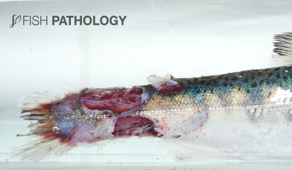
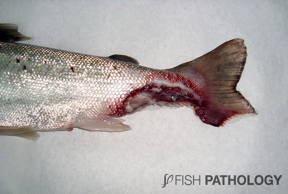
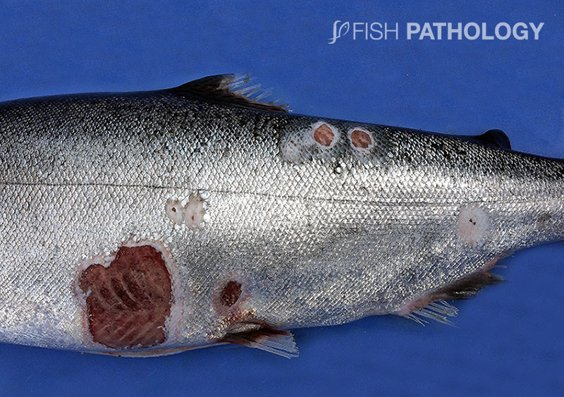
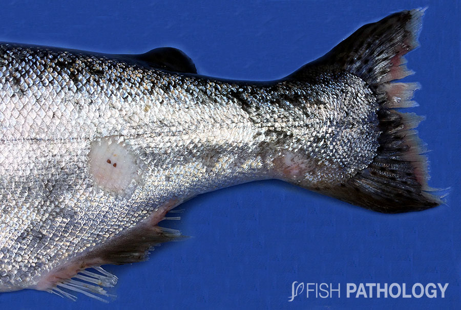
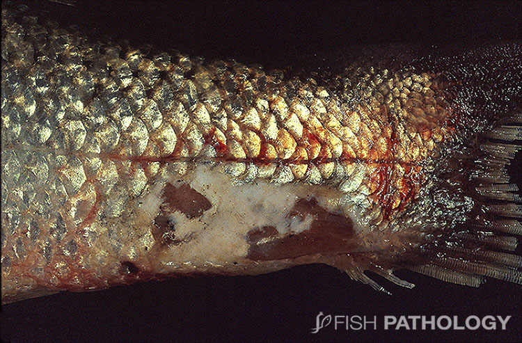
Affected fish may have an eroded mouth, frayed fins, tail rot, or characteristic dark necrotic patches on the skin surface.
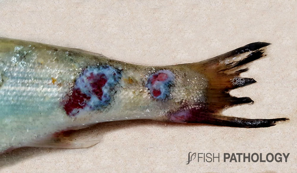
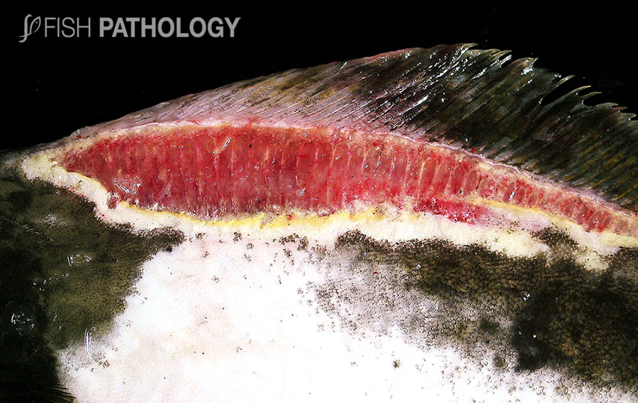
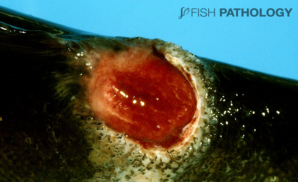
Skin lesions are often haemorrhagic and surrounded by a wider zone of darkened dermis, representing activated dermal melanocytes, or by a zone of yellow, representing the yellow-pigmented bacteria themselves.
Epidermal erosion with attached bacterial mats often progresses to low level inflammation in scale pockets, with oedema, and often scale loss, followed by full-thickness epidermal erosion and haemorrhagic ulceration; these latter lesions can have a prominent white rim of exposed collagen.
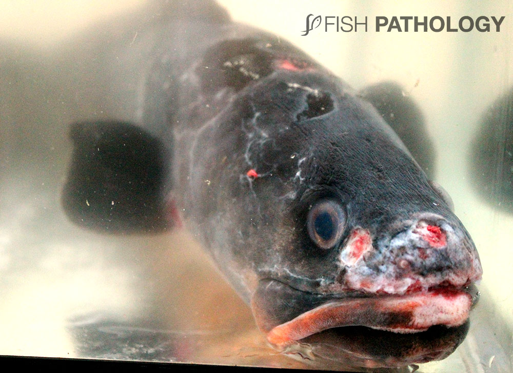
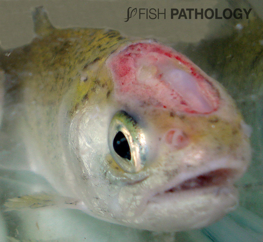
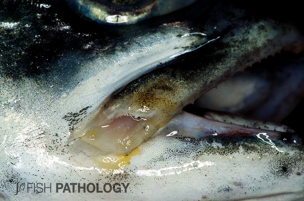
Lesions on the head often involve the mouth and include focal yellow bacterial mats on the palate and base of the teeth.
The lesions range from small and hardly visible to multiple and plaque-like, with erosion of the upper and/or lower jaw in severe cases.
Even small lesions can be significant, as these bacteria are highly toxigenic and can have profound systemic consequences.
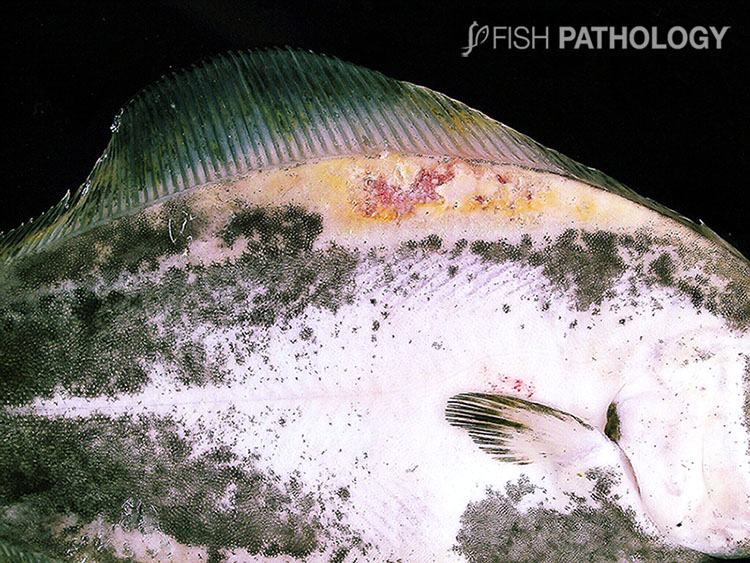
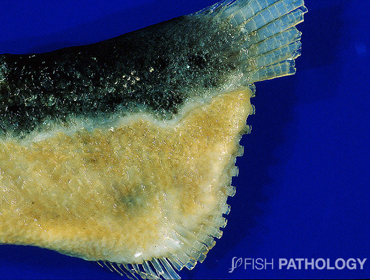
Figure 10: Severe ulcerative dermatitis in farmed halibut associated with filamentous bacteria, likely T. maritimum. The lesion is bright yellow, due to the bacteria.
In severe outbreaks, erosion down to the skull, with exposure of bone may be found. Internal lesions include ascites, paleness and swelling of liver, spleen and stomach. The presentation can suggest toxaemia.
Lesions in gills can be found anywhere, including the gill arches, but they are generally restricted to lamellae and filaments and include severe necrosis and even infarction.
Some jellyfish have been shown to carry the bacteria and it has been suggested that they may act as reservoirs of infection and even vectors, especially those species of jellyfish small enough to pass through the mesh of nets, and hence also be present in the water passing over the gills.
The precise relationship between the bacteria and the jellyfish is unclear; are they a pathogen of the jellyfish, or are they present in a symbiotic or commensal capacity?
REFERENCES
- Avendaño‐Herrera, R., Irgang, R., Sandoval, C., Moreno‐Lira, P., Houel, A., Duchaud, E., … & Ilardi, P. (2016). Isolation, characterization and virulence potential of Tenacibaculum dicentrarchi in salmonid cultures in Chile. Transboundary and emerging diseases, 63(2), 121-126.
- Avendaño-Herrera, R., Toranzo, A. E., & Magariños, B. (2006). Tenacibaculosis infection in marine fish caused by Tenacibaculum maritimum: a review. Diseases of aquatic organisms, 71(3), 255-266.
- Ferguson, H. W., Christian, M. D., Hay, S., Nicolson, J., Sutherland, D., & Crumlish, M. (2010). Jellyfish as vectors of bacterial disease for farmed salmon (Salmo salar). Journal of veterinary diagnostic investigation, 22(3), 376-382.
- Gourzioti, E., Kolygas, M. N., Athanassopoulou, F., & Babili, V. (2016). Tenacibaculosis in aquaculture farmed marine fish. Journal of the Hellenic Veterinary Medical Society, 67(1), 21-32.
- Grothusen, H., Castillo, A., Henríquez, P., Navas, E., Bohle, H., Araya, C., … & Mancilla, M. (2016). First complete genome sequence of Tenacibaculum dicentrarchi, an emerging bacterial pathogen of salmonids. Genome Announc., 4(1), e01756-15.
- Haridy, M., Hasheim, M., El-Galil, M. A., Sakai, H., & Yanai, T. (2015). Pathological Findings of Tenacibaculum maritimus Infection in Black Damselfish, Neoglyphieodon melas and Picasso Triggerfish, Rhinecanthus assasi in Red Sea, Egypt. Veterinary Science & Technology, 6(2), 1.
- Roberts, Ronald J. (2012). Fish Pathology. Hoboken, NJ: Wiley-Blackwell
