
Aquaculture of cobia (Rachycentrum canadum) has gained popularity in the last decade, and this species is now farmed in several countries in Latin America and Asia.
Epitheliocystis is a chlamydia or rickettsia-like organism infecting the gills and skin of a variety of species in both fresh and saltwater. The disease has been reported in at least 90 species of marine and freshwater fish in both the southern and northern hemispheres.
At least four bacterial species may be involved in this condition: Candidatus Piscichlamydia salmonis (Salvelinus alpinus), Candidatus Clavochlamydia salmonicola (Salmo salar, salmo trutta), Candidatus Branchiomonas cisticola (Salmo salar), and Endozoicomonas elysicola (Rachycentrum canadum).
It is a common disease of fish characterized by
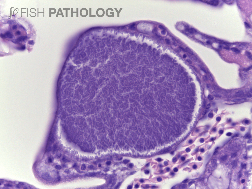
the presence of cyst-like basophilic inclusions in the skin and gills. Although
in wild fish epitheliocystis is usually a benign disease, in aquaculture-reared
fish it can produce high mortalities, especially in infections during early life
stages.
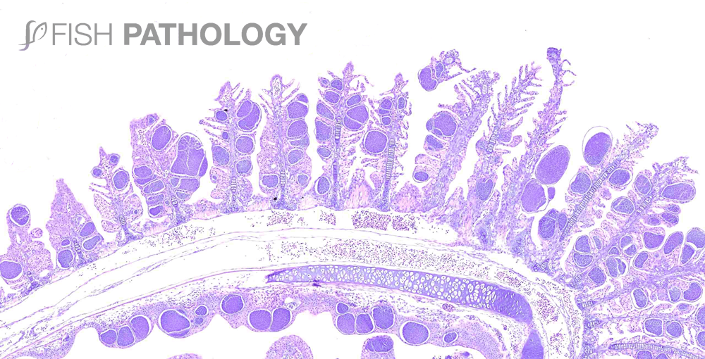
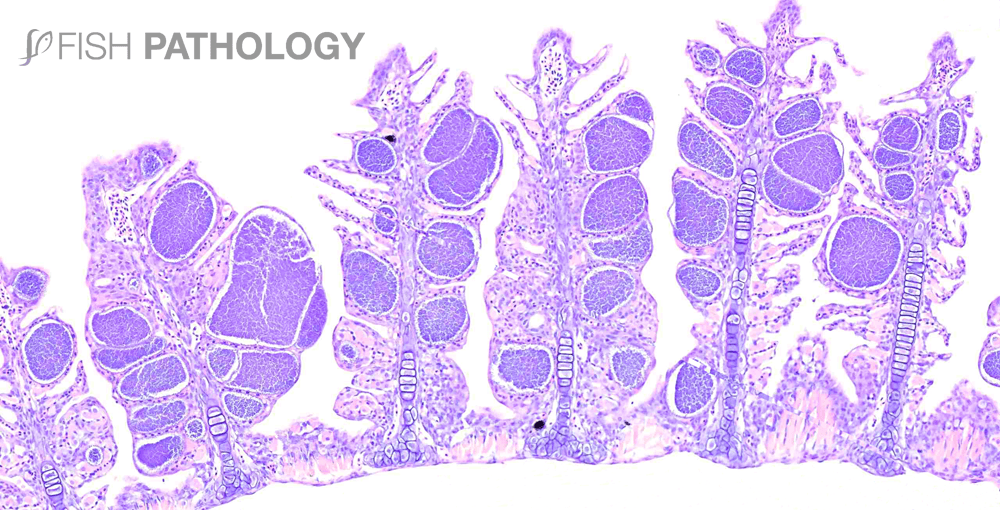
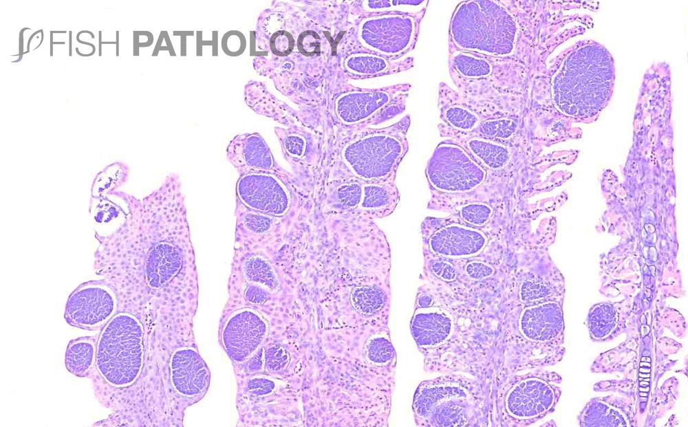
In Cobia, histopathological examination of larvae can show the presence of a large number of dense basophilic bodies (cysts), located within the hypertrophied epithelial cells of gills. Cysts are located predominantly at the base of the filaments with fewer cysts detected near the distal area. However, in severe infections cysts can be present throughout the entire length.
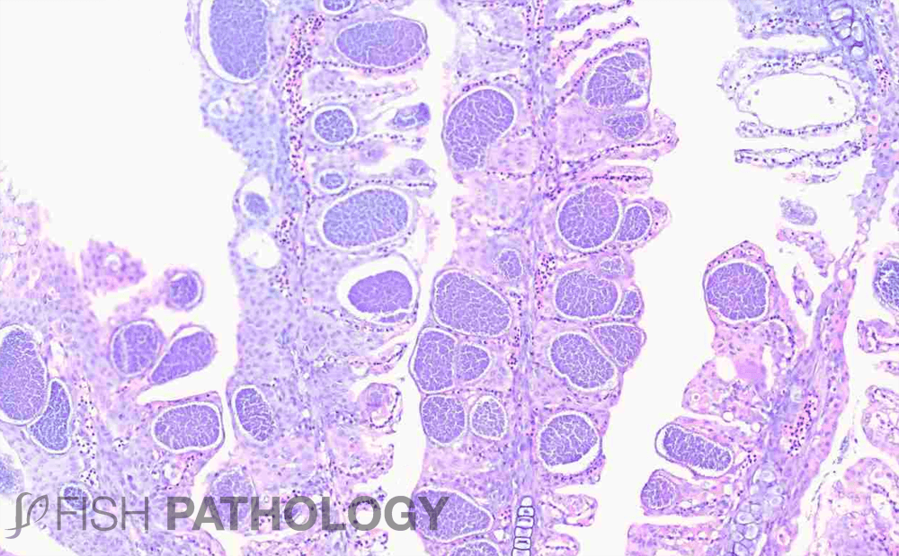
Pathological changes centre round respiratory disturbances, brought about by the space-occupying effects of so many large cysts which are often accompanied by severe epithelial hyperplasia, inflammation in sub-epithelial and epithelial tissue, mucus cell hyperplasia with fusion of lamellae, telangiectasia and infiltration of macrophages.
REFERENCES
- Blandford, M. I., Taylor‐Brown, A., Schlacher, T. A., Nowak, B., & Polkinghorne, A. (2018). Epitheliocystis in fish: an emerging aquaculture disease with a global impact. Transboundary and emerging diseases, 65(6), 1436-1446.
- Bruno, D.W, 2013, “A Colour Atlas of Salmonid Diseases”, New York-London, Springer
- Mendoza, M., Güiza, L., Martinez, X., Caraballo, X., Rojas, J., Aranguren, L. F., & Salazar, M. (2013). A novel agent (Endozoicomonas elysicola) responsible for epitheliocystis in cobia Rachycentrum canadum larvae. Diseases of aquatic organisms, 106(1), 31-37.
- Noga, E.J, 2000, “Fish Diseases, diagnosis and treatment”, Iowa State University Press.
