
Strawberry Disease (SD) is a chronic, nonlethal skin condition that affects Rainbow Trout (Oncorhynchus mykiss) in the United States, several European countries (Scotland, England, Switzerland, Germany, France, among others), Chile and Peru (Sandoval 2017, unpublished report). In Europe it is also known as Red Mark Syndrome (RMS) or Cold-Water Strawberry Disease (CWSD).
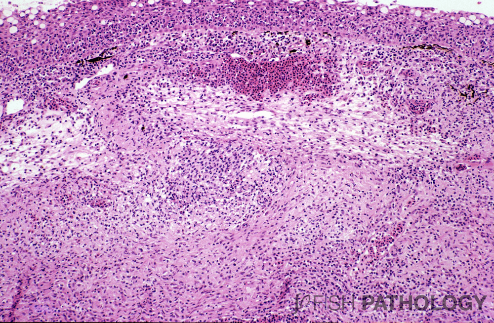
SD is characterized by raised, sometimes bright red inflammatory lesions that can occur in almost any size of fish bigger than fingerlings, but usually in growing or market-sized fish. The disease presents as a severe, full-thickness, largely non-ulcerating, dermatitis centred on the dermis, with an inflammatory response dominated by small mononuclear cells. The severity of the response is such as to suggest a hypersensitivity reaction.

In both moderate and severe cases, scale resorption is observed concomitant with osteoclastic activity and oedema-induced distention of the scale pocket. In mild lesions, lymphocyte infiltration is observed in the stratum spongiosum and around the scale pocket; in moderate cases, marked oedema can be observed, often accompanied by mild neutrophil infiltration. In advanced cases, scales are usually absent, having been completely resorbed by osteoclasts, but the inflammatory response can extend down into the sub-cutis, or even the muscle layer, sometimes accompanied by myonecrosis.
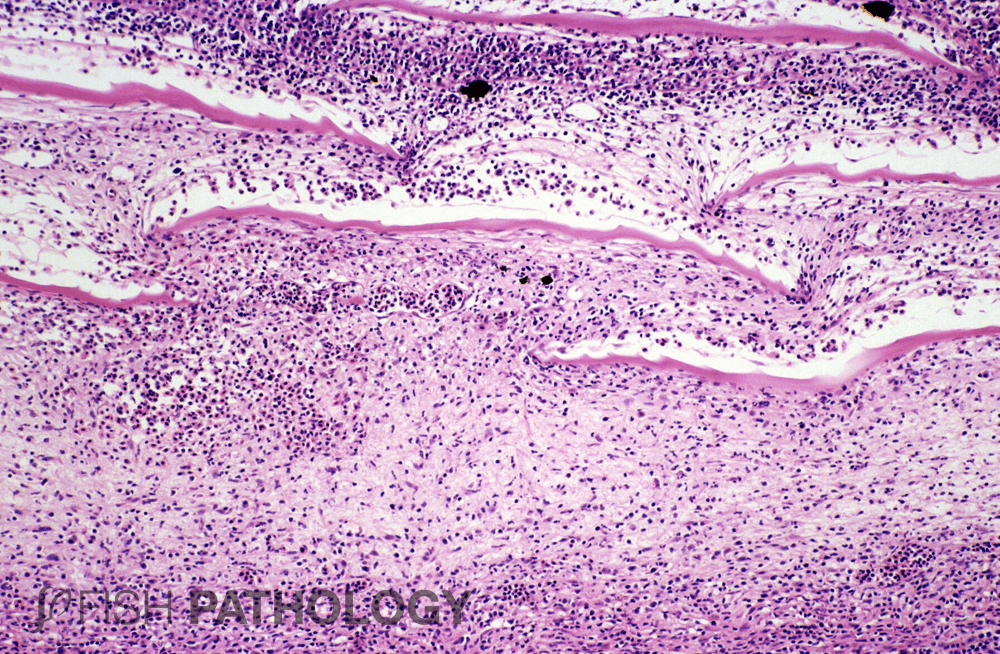
The epidermis is largely unaffected in mild or moderate cases, but erosion and ulceration of the epidermis are observed in severe cases, along with mild lymphocytic intra-epithelial infiltration. If the skin is indeed ulcerated, interpretation of the lesions can be hampered by secondary infection. In some severe cases, mild mucous and epidermal hyperplasia are also seen. In moderate to severe cases, collagen fibers of the dermis are infiltrated by lymphocytes and some plasmacytes.
REFERENCES
- Lloyd, S. J., LaPatra, S. E., Snekvik, K. R., St-Hilaire, S., Cain, K. D., & Call, D. R. (2008). Strawberry disease lesions in rainbow trout from southern Idaho are associated with DNA from a Rickettsia-like organism. Diseases of aquatic organisms, 82(2), 111-118.
- Sandoval, C., Infante, J., Abad, J., Ferguson, H. W., Paredes, E., Valdebenito, S., … & Avendaño-Herrera, R. (2016). Case report: Strawberry disease in farmed Chilean rainbow trout. Journal of aquatic animal health, 28(1), 1-10.
- Verner-Jeffreys, D. W., Pond, M. J., Peeler, E. J., Rimmer, G. S. E., Oidtmann, B., Way, K., … & Feist, S. W. (2008). Emergence of cold water strawberry disease of rainbow trout Oncorynchus mykiss in England and Wales: outbreak investigations and transmission studies. Diseases of aquatic organisms, 79(3), 207-218.
- Ferguson, H. W., Girons, A., Rizgalla, G., LaPatra, S., Branson, E. J., Mackenzie, K., … & Crumlish, M. (2006). Strawberry disease in rainbow trout in Scotland: pathology and association with Flavobacterium psychrophilum.
