
Cataracts are changes in the clarity of the lens so that it appears cloudy. Such changes are a clinical or gross diagnosis, and there may be no histopathological correlate.
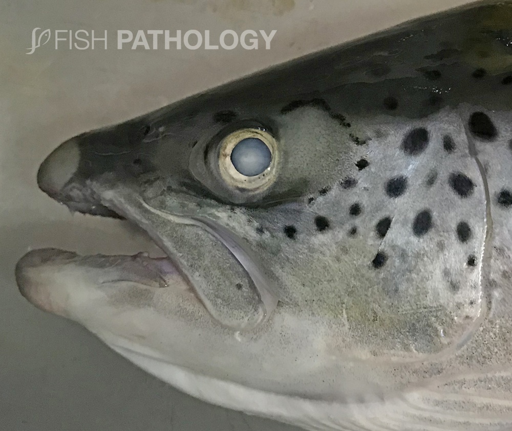
Nevertheless, opacity of the lens results in reduced ability to see.

Cataracts may be divided into primary or secondary disease. In primary cataract, opacity of the lens is the only sign of disease in the eye, while in secondary cataract the lens changes occur as a consequence of other intraocular disease.
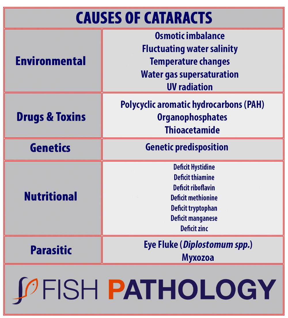
A wide variety of causes of primary cataracts have been described in farmed fish. These include nutritional imbalances, rapid growth, variation in water temperature, exposure to ultraviolet light, gas supersaturation, change in water salinity, genetic susceptibility, environmental toxins, toxic side effects of drug treatment, and parasitic infestation.

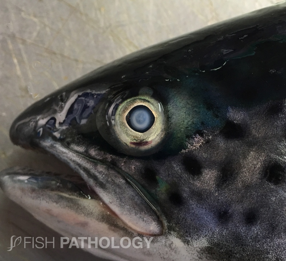
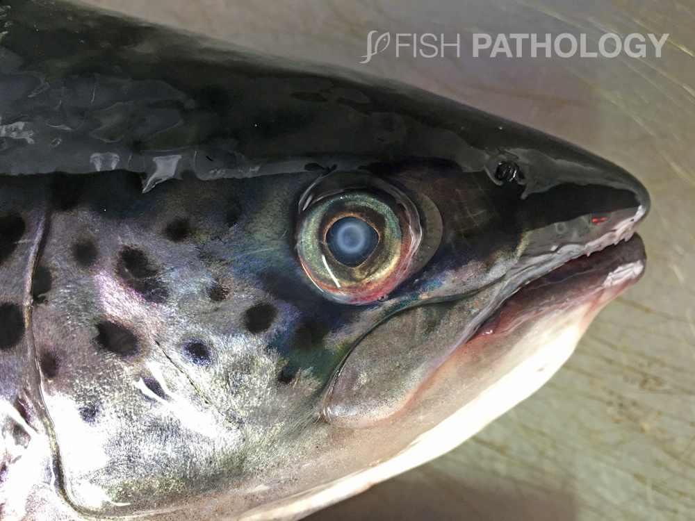
Grossly, cataracts are usually observed as a partial or total opacity of the lens. In initial stages small white spots are observed in the central part of the lens and in advanced stages the entire lens is involved. Sometimes white halos are observed in the lens.
These are typically posterior cataracts and have been associated with nutritional deficiencies.
REFERENCES
- Bjerkås, E., Waagbø, R., Sveier, H., Breck, O., Bjerkås, I., Bjørnestad, E., & Maage, A. (1996). Cataract development in Atlantic salmon (Salmo salar L) in fresh water. Acta Veterinaria Scandinavica, 37(3), 351-360.
- Remø, S. C., Hevrøy, E. M., Breck, O., Olsvik, P. A., & Waagbø, R. (2017). Lens metabolomic profiling as a tool to understand cataractogenesis in Atlantic salmon and rainbow trout reared at optimum and high temperature. PloS one, 12(4).
- Ersdal, C., Midtlyng, P. J., & Jarp, J. (2001). An epidemiological study of cataracts in seawater farmed Atlantic salmon Salmo salar. Diseases of Aquatic organisms, 45(3), 229-236.
- Ferguson, H. W., 2006, Systemic Pathology of Fish, London, UK, Scottian Press.
- Wall, T., & Bjerkas, E. (1999). A simplified method of scoring cataracts in fish. Bulletin of the European Association of Fish Pathologists, 19(4), 162-165.
