Figure 1. Multiple giant cells in the peritoneal cavity of salmon, a response to vaccine, its bright red character apparent (arrows). Similar to so-called Splendore-Hoeppli reactions (asteroid bodies).

GIANT CELLS IN FISH

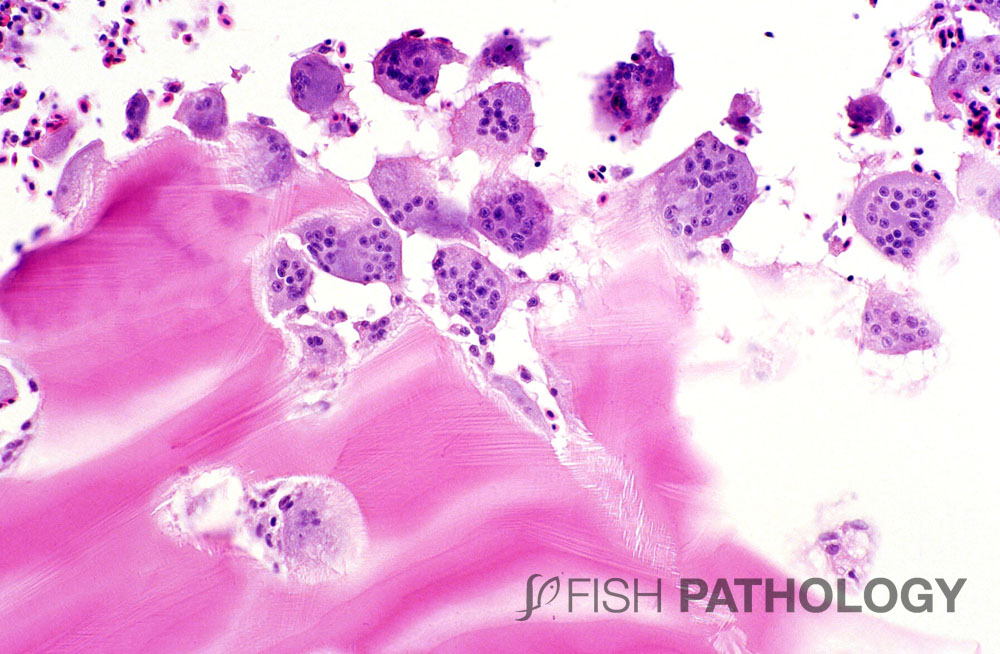
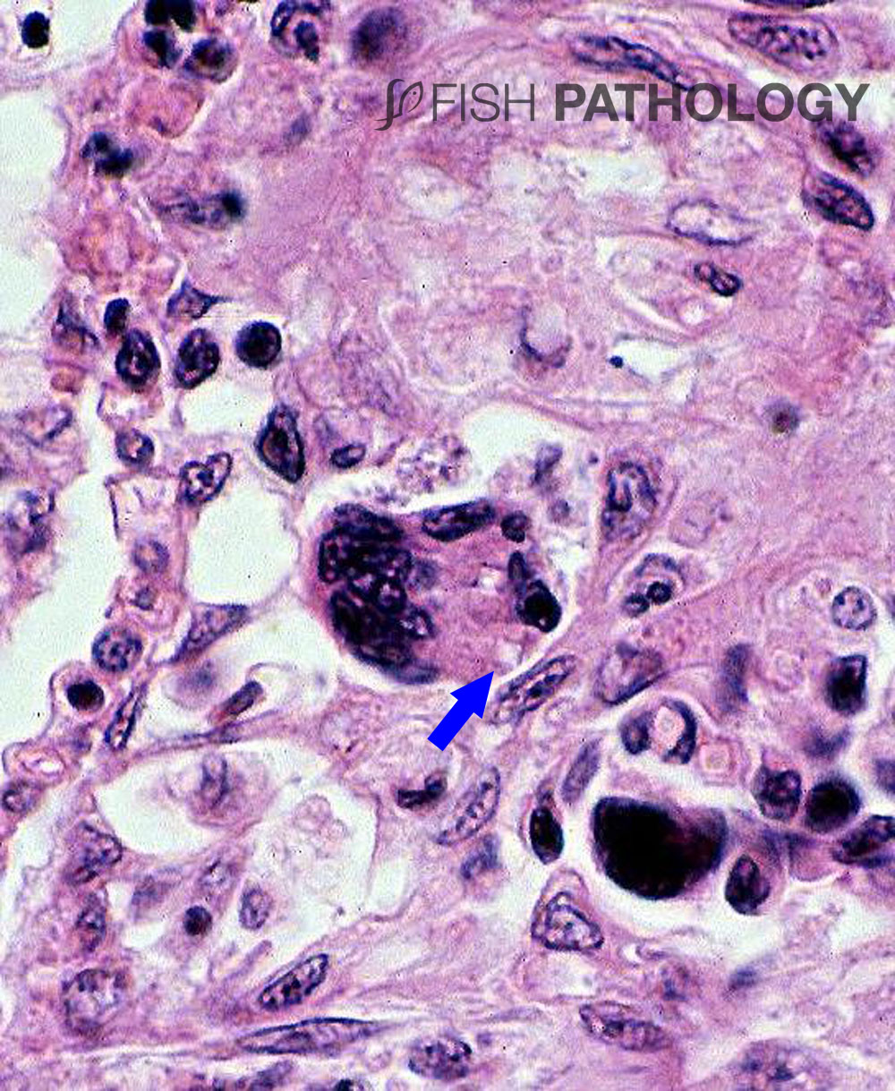
Multinucleated cells, often simply called giant cells, are found in a variety of situations in teleost fish.

Figure 1. Atlantic salmon. Multiple giant cells in the peritoneal cavity of salmon, a response to vaccine, its bright red character apparent (arrows). Similar to so-called Splendore-Hoeppli reactions (asteroid bodies).

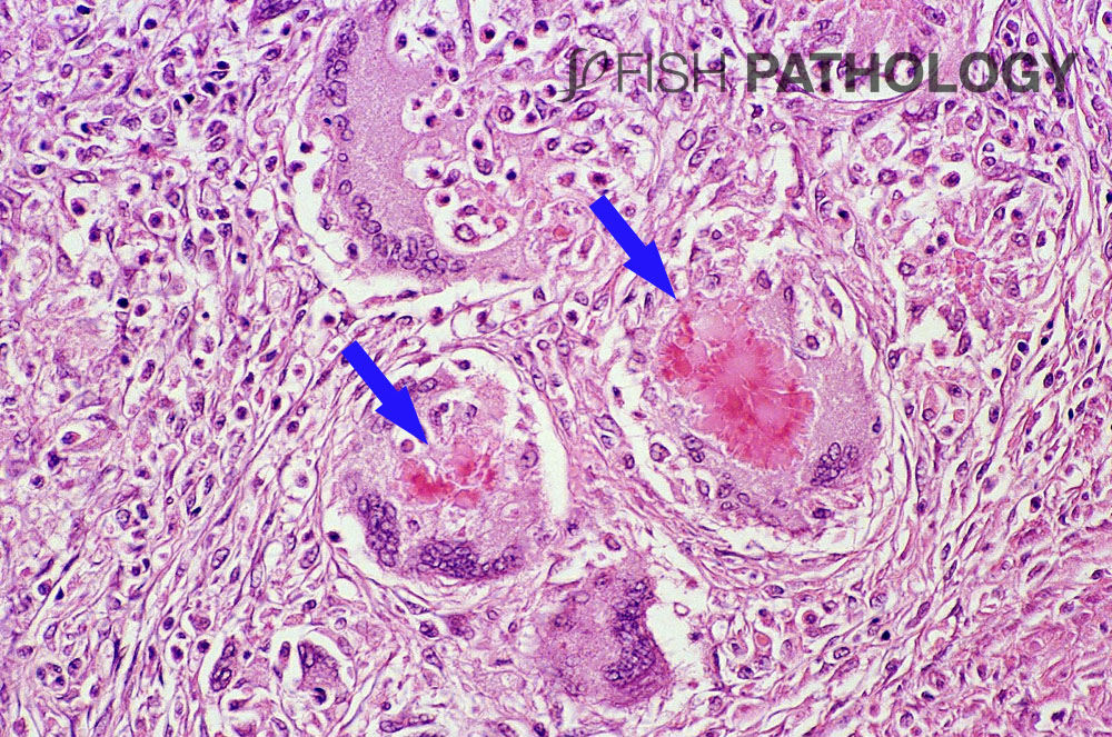
They are not uncommon in granulomatous inflammatory responses, such as bacterial kidney disease, proliferative kidney disease, or as a response to vaccine, and are a result of fusion of macrophages or epithelioid cells.

Figure 2. Osteoclastic remodelling of dermal scale in this example of dermatitis in trout. Note the “scoops” of bone in which the osteoclasts have been actively dissolving the scale. Oedema seems to be a trigger for osteoclasis, maybe due to hypoxia – bone has high oxygen requirements
Figure 2. Osteoclastic remodelling of dermal scale in this example of dermatitis in trout. Note the “scoops” of bone in which the osteoclasts have been actively dissolving the scale. Oedema seems to be a trigger for osteoclasis, maybe due to hypoxia – bone has high oxygen requirements.


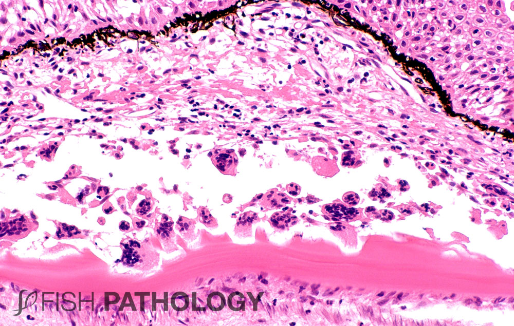
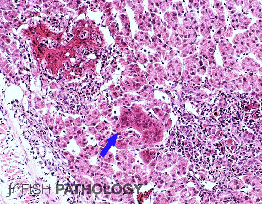
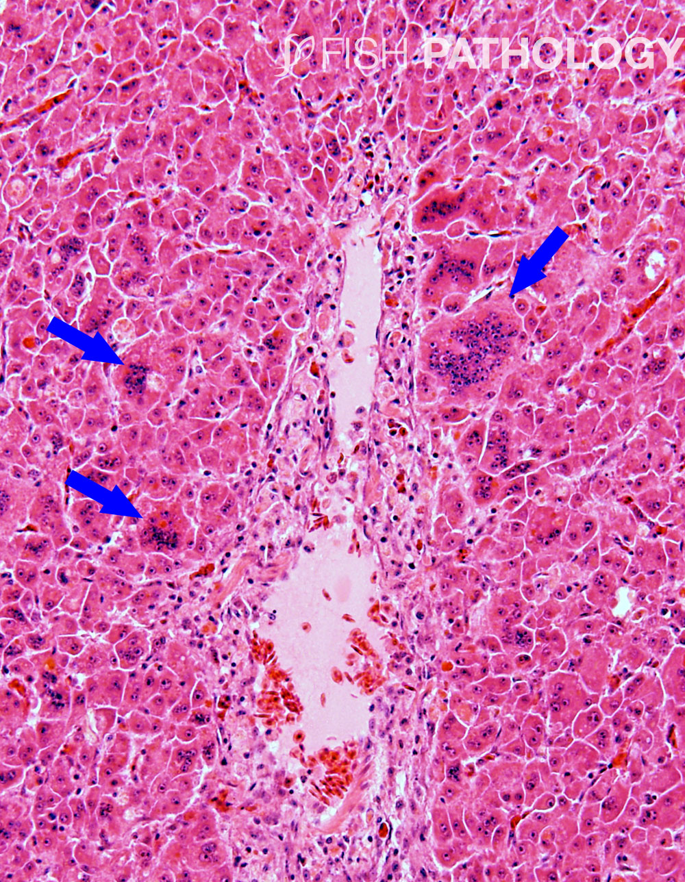
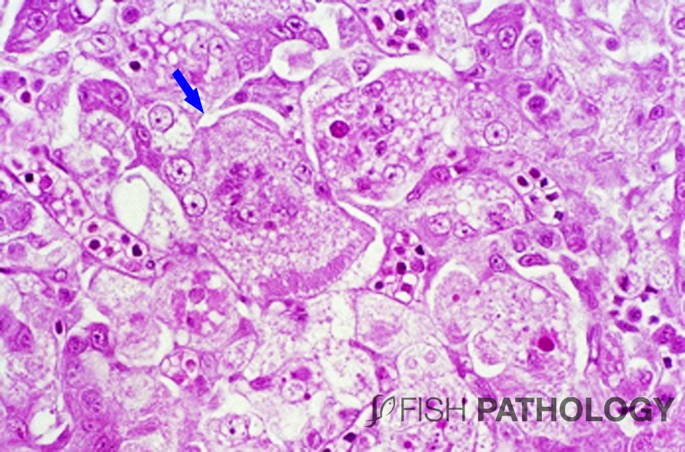
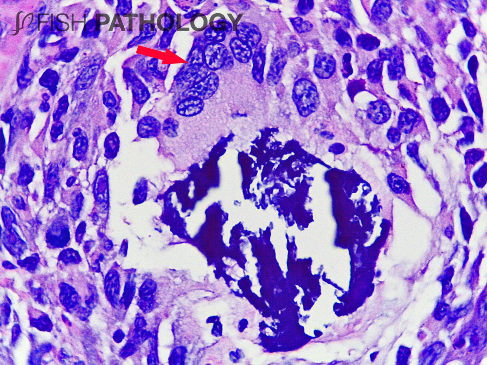
They are also found in virus infections, so-called syncytial giant cells, in which a number of non-macrophage cells, such as hepatocytes, fuse together. But giant cells are also found in normal fish as osteoclasts.

Figure 3. Foreign-body type giant cells, part of a granulomatous response to metazoan eggs, one of which can be seen in the process of being engulfed by the giant cell (arrow). Chitin, found in metazoa (and some fungi), is hard to digest, hence the need for giant cells.


Although osteoclasts in fish can be mononuclear, the multinucleated ones are more easily seen histologically.

Figure 4. Reoviral hepatitis in juvenile halibut. A few syncytia are present (arrow), as is a large focus of caseonecrotic liver, and a focally extensive area of inflammation.
Figure 5. Osteoclastic remodelling of the axial skeleton in a goldfish.


These cells are responsible for removing or remodelling damaged tissue or for resorbing bone, as seen during periods of high calcium demand, for example at spawning time.

Figure 6. Orthomyxoviral hepatitis in juvenile tilapia – “syncytial hepatitis”. Several syncytial giant cells are present (arrows).


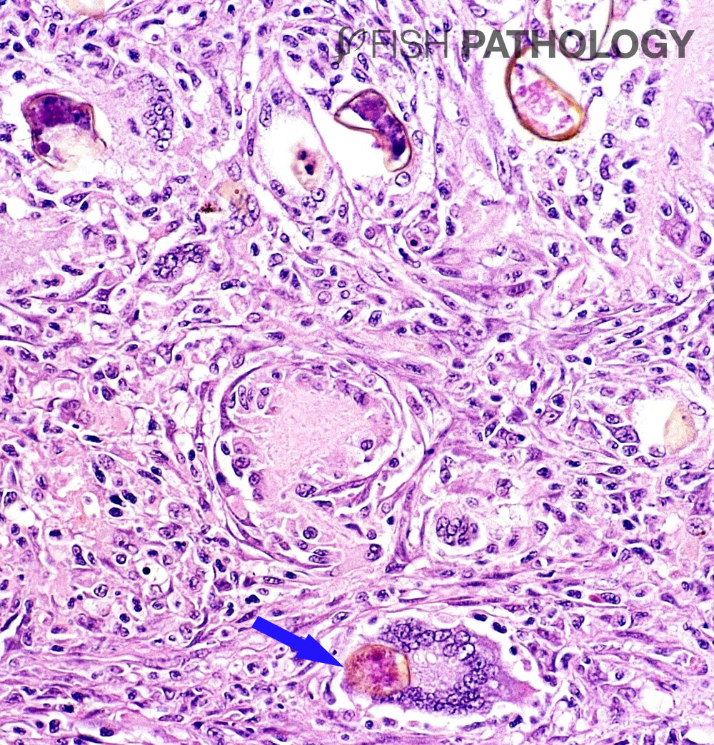
In skin biopsies, it is important to differentiate osteoclasts removing scales damaged, for example in dermatitis, from foreign-body type giant cells, although both can occur at the same time.

Figure 7. Liver from rainbow trout with “cardio-hepatic syndrome” showing several very large multi-nucleated hepatocytes that have a markedly vacuolated cytoplasm. This rare disease is associated with intra-nuclear herpes-like virus.
Figure 8. Kidney from Atlantic salmon in sea water, with nephrocalcinosis. Note the giant cell engulfing calcium (arrow).
Figure 9. Gill from coho salmon exposed to diatoms, showing cross-sections of setae (arrow) within the cytoplasm of giant cell
Figure 9. Gill from Coho salmon exposed to diatoms, showing cross-sections of setae (arrow) within the cytoplasm of giant cell.


REFERENCES

  • Couso, N., Castro, R., Noya, M., Obach, A., & Lamas, J. (2002). Formation of short‐lived multinucleated giant cells (MGCS) from cultured gilthead seabream macrophages. The Anatomical Record: An Official Publication of the American Association of Anatomists267(3), 204-212.
  • Goodwin, A. E., & Grizzle, J. M. (1991). Granulomatous inflammation and monstrous giant cells in response to intraperitoneal hormone implants in channel catfish (Ictalurus punctatus). Journal of comparative pathology104(2), 147-160.
  • Speare, D. J., Brackett, J., & Ferguson, H. W. (1989). Sequential pathology of the gills of coho salmon with a combined diatom and microsporidian gill infection. The Canadian Veterinary Journal30(7), 571.

2 Comments

  1. Paola

    Very nice and interesting pathological events. I would like to point out that even during Exophiala infection in trout, multinucleated cells are very common.
    Thanks for very good work
    Paola

totop