Rainbow trout with PKD. There is a marked hyperplasia of interstitium with the kidney thrown into bulbous ridges. The reddening is tge result of secondary yersinia infection.

Proliferative Kidney Disease in Salmonids (PKD) – Gross Pathology

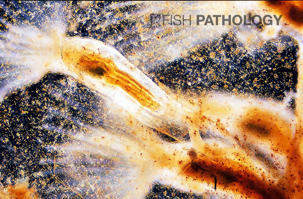
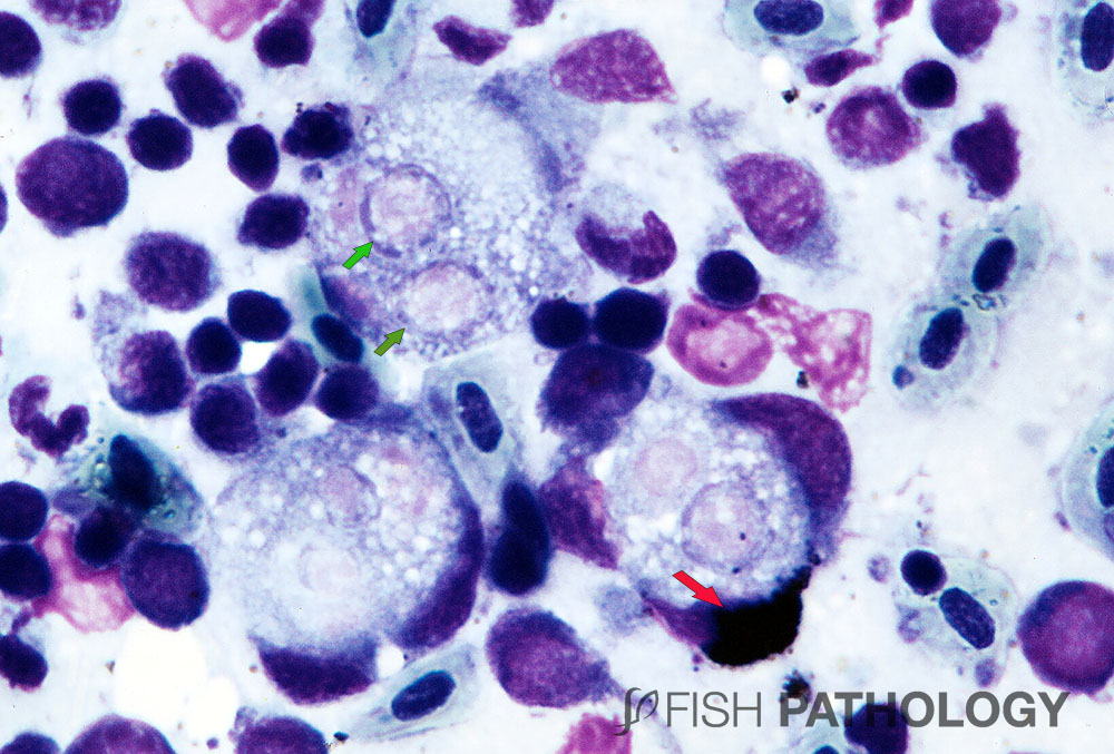
Proliferative kidney disease (PKD) is an endoparasitic disease of salmonid fish caused by Tetracapsuloides bryosalmonae (Myxozoa: Malacosporea).  This chronic, largely renal interstitial disease is caused by the extraporogonic but intracellular stages of the parasite, which cause a severe granulomatous host response.

Rainbow trout with PKD. There is a marked hyperplasia of interstitium with the kidney thrown  into bulbous ridges. The reddening is tge result of secondary  Yersinia infection.
Figure 1. Rainbow trout with PKD. There is a marked hyperplasia of interstitium with the kidney thrown into bulbous ridges. The reddening is the result of secondary Yersinia infection.

The severity of the disease is linked to water temperature, with roughly 15 degrees °C as the cut-off: below that temperature, lesions and clinical disease are minimal.

These bryozoa are infected with T. bryosalmonae.
Figure 2. These bryozoa are infected with T. bryosalmonae.


Above that temperature, however, lesions can be severe and mortality high. Inevitably, global warming has resulted in spread of the disease.

Rainbow trout showing severe PKD. Note that the entire kidney has been thrown into grey bulbous ridges, a consequence of severe granulomatous inflammation.
Figure 3. Rainbow trout showing severe PKD. Note that the entire kidney has been thrown into grey bulbous ridges, a consequence of severe granulomatous inflammation.


All ages of fish are susceptible unless they have been previously exposed to the parasite: thus, immunity is possible. Similarly, some strains of fish are more resistant than others. Filter-feeding bryozoa (also known as moss animals) have been shown to be responsible for releasing T. bryosalmonae spores, even a small number of which can then infect fish and lead to PKD.

Giemsa-stained impression smear from kidney of rainbow trout with PKD showing developing (presporogonic) stages of Tetracapsuloides bryosalmonae (green arrows) within host cells. Note that the host is mounting an immune response, as evidenced by the presence of surrounding macrophages, one of which is a melano-macrophage (red  arrow).
Figure 4. Giemsa-stained impression smear from kidney of Rainbow trout with PKD showing developing (presporogonic) stages of Tetracapsuloides bryosalmonae (green arrows) within host cells. Note that the host is mounting an immune response, as evidenced by the presence of surrounding macrophages, one of which is a melano-macrophage (red arrow).


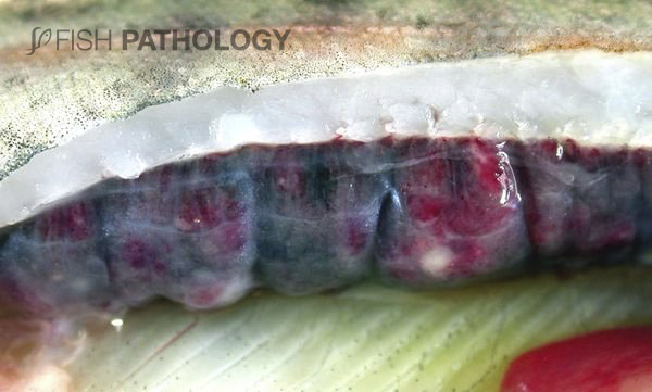
Fish with PKD are often lethargic and darker than normal. As the primary target organ, the kidney may become so massively enlarged as to be visible externally along the lateral line. Exophthalmia, ascites and anaemia are other gross features, probably the results of the disease on vascular integrity, leading to haemorrhage and reduced haematopoiesis due to the granulomatous lesions (myelophthisic anaemia?).

Chinook salmon showing abdominal distension and exophthalmia. Although these fish have BKD, these grossly visible signs are characteristic for fish with PKD.
Figure 5. Chinook salmon showing abdominal distension and exophthalmia. Although these fish have BKD, these grossly visible signs are characteristic for fish with PKD.


Both anterior and posterior portions of the kidney may be thrown into grey bulbous ridges or discrete nodules, depending on the severity of its involvement. Splenomegaly is usually present and indeed may be one of the earliest indications of infection.


REFERENCES

  • El‐Matbouli, M., & Hoffmann, R. W. (2002). Influence of water quality on the outbreak of proliferative kidney disease–field studies and exposure experiments. Journal of Fish Diseases25(8), 459-467.
  • Ferguson, H.W., 2006, Systemic Pathology of Fish, London, UK, Scotian Press.
  • Palikova, M., Papezikova, I., Markova, Z., Navratil, S., Mares, J., Mares, L., … & Schmidt-Posthaus, H. (2017). Proliferative kidney disease in rainbow trout (Oncorhynchus mykiss) under intensive breeding conditions: Pathogenesis and haematological and immune parameters. Veterinary parasitology238, 5-16.
  • Wahli, T., Knuesel, R., Bernet, D., Segner, H., Pugovkin, D., Burkhardt‐Holm, P., … & Schmidt‐Posthaus, H. (2002). Proliferative kidney disease in Switzerland: current state of knowledge. Journal of Fish Diseases25(8), 491-500.

By: Hugh Ferguson

Dr Ferguson earned his veterinary degree from the Royal (Dick) School of Veterinary Studies, Edinburgh, Scotland and held a Wellcome Research Fellowship at the Institute of Aquaculture, Stirling University where he obtained his PhD. He then worked for 4 years as a diagnostic pathologist at the Veterinary Research Laboratories, Belfast, Northern Ireland, prior to moving to Canada. He left Ontario Veterinary College after 19 years as a full professor of veterinary pathology, to return to Scotland to become head of diagnostic pathology in Stirling. During all this time he became board-certified in the American college of veterinary pathology (ACVP), and a Fellow of the Royal College of Pathologists (FRCPath, London). After Scotland he moved to become chair of veterinary pathology, and Senior Research Fellow in Windward Islands Research and Education Foundation (WINDREF), St George’s University (SGU), Grenada, West Indies. He has published more than 230 papers in refereed journals.

totop