
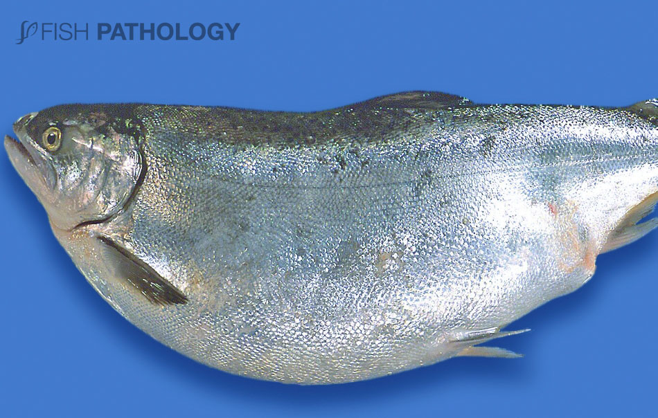
Bloat is a non-infectious condition of salmonids where the abdomen is abnormally distended by an enlarged, fluid-filled stomach. The wall of the enlarged stomach wall is thin and flaccid. Occasionally the swimbladder is also affected.
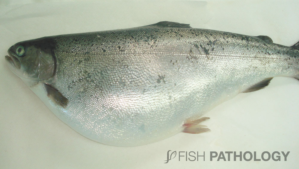
The condition is seen in salmonids reared in sea water and fed fishmeal-based pelleted rations. While it has been reported occasionally in Atlantic salmon (Salmo salar L.) members of the genus Oncorhynchus are more susceptible. These include Rainbow trout (O. mykiss), Chinook salmon (O. tshawytscha), and Coho salmon (O. kisutch).

The pathophysiology of GDAS (gastric dilation and air sacculitis) or colloquially called “Bloat” is not completely clear. The condition involves a combination of a failure of osmotic regulation and nutrient overloading of the gastro-intestinal tract.

Farmed salmon fed nutrient-rich rations are likely to develop some degree of permanent stomach distention. Once this has occurred, factors such as changes in the composition or properties of the food, alteration of the feeding regime, increased stress (e.g. from handling or predation) or environmental changes (e.g. increased water temperatures, low water oxygen levels) may lead to the development of bloat.
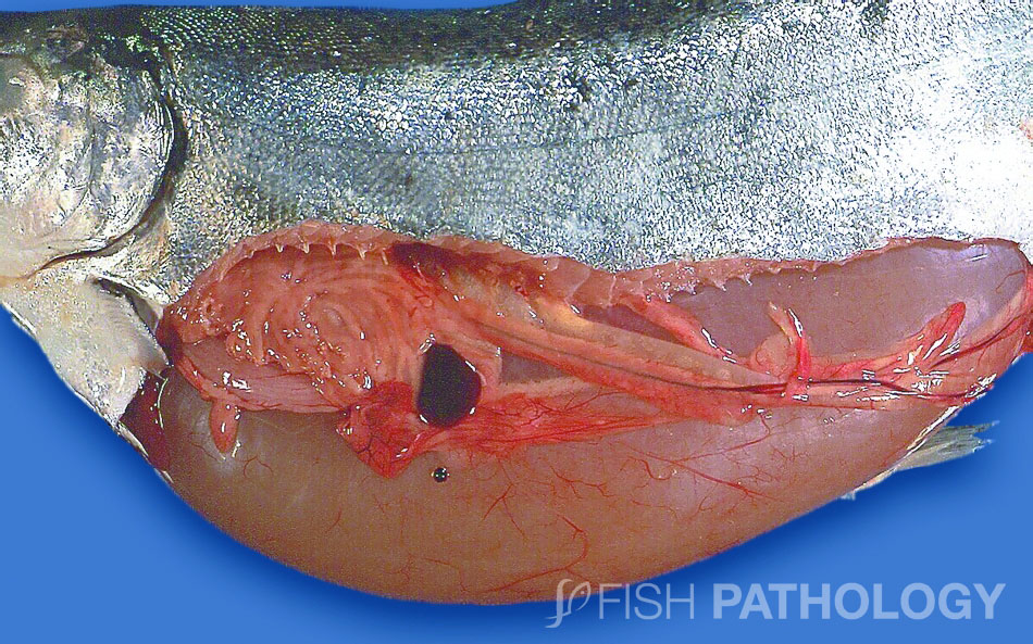
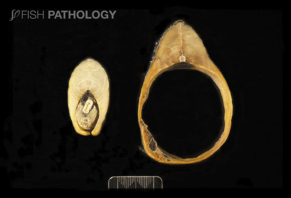
The distended thin-walled stomach of
bloat-affected salmonids contains an excessive amount of liquid and variable
amounts of food and oil that are often regurgitated onto the water surface,
usually the first symptom of the condition.
The luminal contents of affected swimbladders are usually contaminated with a mixed microbial flora and in chronic cases, the swimbladder wall may become severely congested and inflamed. Stomach and swimbladder dilation may occur either singly or together.
The thinning of the abdominal wall musculature, absence of contraction of the stomach wall and reduction in abdominal fat are consistent with prolonged distention of the abdomen, reduced energy intake and stress.
Farm experience has shown that bloat in salmonids can be controlled by reducing food intake, altering the composition of the diet and using appropriate strategies to reduce stress caused by digestion and gastric emptying.
REFERENCES
- Anderson, C. D. (2006). A review of causal factors and control measures for bloat in farmed salmonids with a suggested mechanism for the development of the condition. Journal of fish diseases, 29(8), 445-453.
- Ferguson, H.W., 2006, Systemic Pathology of Fish, London, UK, Scotian Press.
- Forgan, L. G., & Forster, M. E. (2007). Development and physiology of gastric dilation air sacculitis in Chinook salmon, Oncorhynchus tshawytscha (Walbaum). Journal of fish diseases, 30(8), 459-469.
- Lumsden, J. S., Wybourne, B., Minamikawa, M., & Tubbs, L. (2010). Gastric dilation and air sacculitis in Chinook salmon, Oncorhynchus tshawytscha (Walbaum); correlation of macroscopic and microscopic lesions, and relationship of the syndrome to glomerulonephritis and serum biochemistry. Journal of fish diseases, 33(9), 737-747.
- Rørvik, K. A., Skjervold, P. O., Fjæra, S. O., & Steien, S. H. (2000). Distended, water‐filled stomach in seawater farmed rainbow trout, Oncorhynchus mykiss (Walbaum), provoked experimentally by osmoregulatory stress. Journal of Fish Diseases, 23(1), 15-18.
