Figure 5. Phialella quadrata attached to gill raker of Atlantic salmon and showing necrosis of epithelium, loss of basement membrane, and underlying dermal haemorrhage. A close inspection of the interface between the jellyfish and the gill epithelium shows tube-like extensions reaching down through the epithelium – nematocysts?

Jellyfish Lesions in Fish – Histopathology

The negative interactions between jellyfish and fish in aquaculture appear to be an increasing problem. This is partly due to increased numbers of jellyfish, associated with global warming, reduced numbers of their predators, and to the intensification of aquaculture operations in many coastal areas worldwide.

Figure 1. Acute ballooning degeneration of skin on top of head of Atlantic salmon fry in freshwater. This lesion was the result of nematocyst damage from Hydra that were growing on the bottom of the raceways: the fry were stung when foraging for feed.

Most reported problems have occurred in marine-farmed salmonids in northwest Europe. Nevertheless, aquaculture operations in other regions such as Asia, North America, and Australia have also been affected.

Figure 2. Atlantic salmon in seawater with acute damage to epithelium of gill raker caused by Phialella quadrata. This 12mm diameter  jellyfish was small enough to be “inhaled” by the fish, after which it was trapped by the rakers.
Figure 2. Atlantic salmon in seawater with acute damage to epithelium of gill raker caused by Phialella quadrata. This 12mm diameter jellyfish was small enough to be “inhaled” by the fish, after which it was trapped by the rakers.


Jellyfish involved are primarily cnidarians i.e. those species with stinging cells – nematocysts. Several species of jellyfish have been previously linked to fish kill events in marine-farmed fish including hydromedusae, siphonophores, scyphozoans, and ctenophores.

Damage to fish may be direct, through stinging of the skin or gills, or it can be indirect, through de-oxygenation of the surrounding water.

Figure 3. Atlantic salmon in seawater showing severe acute damage to epithelium on gill arch caused by Phialella quadrata. Note the spongy change to epidermis, the superficial layers of which are sloughing, and the dermal inflammation. The basement membrane of the epidermis is brightly eosinophilic, and looks to be degraded (necrobiosis). Has complement been activated?

If the species of jellyfish involved is small, it can be washed through the mesh of nets, and even “inhaled” by the fish, thereby damaging the gills. Loose nematocysts from net washing can lead to similar problems. Nematocysts retain their ability to sting long after the jellyfish is dead.  

Figure 4. Atlantic salmon in seawater showing several cross sections of jellyfish sitting on or close to the surface of the gill arch. Note the necrobiosis of the basement membrane and the dermal oedema.


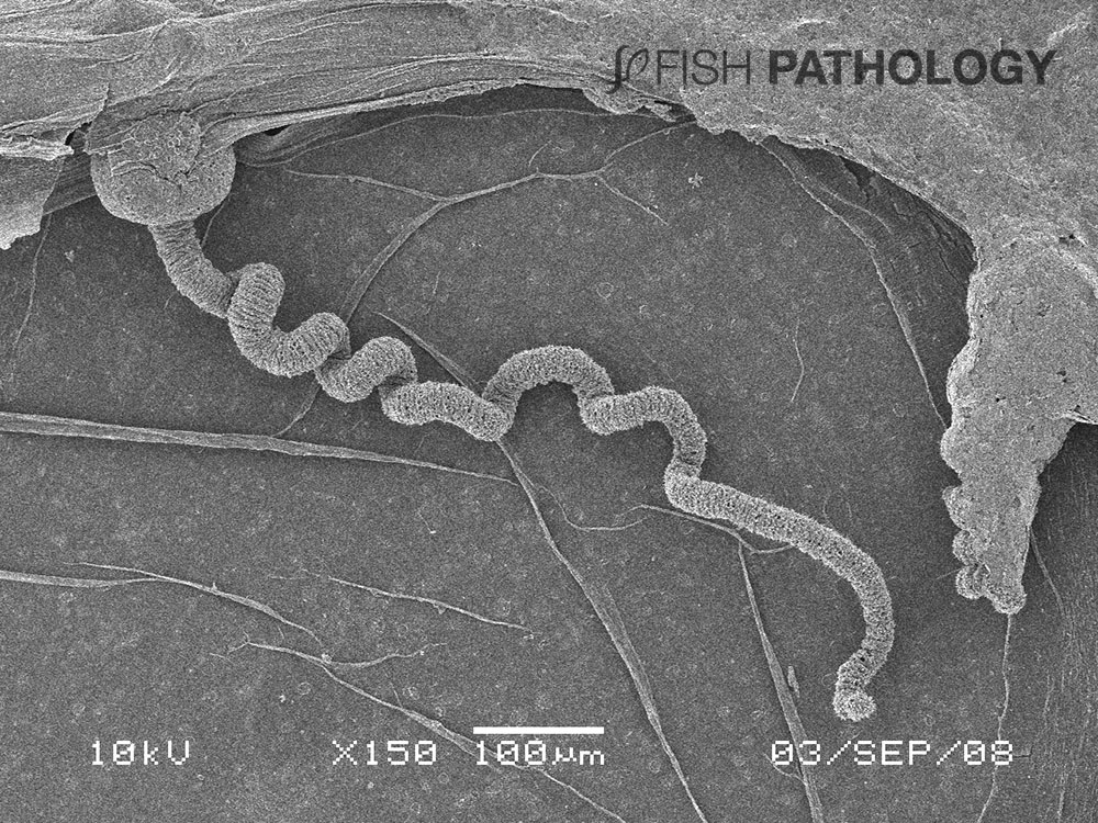
Almost all of the initial gill damage caused by cnidarians is due to their stings. Cnidarian jellyfish are characterized by having millions of microscopic stinging cells, primarily in their tentacles. Inside the cell is a specialized stinging capsule called a nematocyst (mostly 10–20 µm long) that contains a coiled, harpoon-like hollow tube often armed with spines. Nematocyst release can be triggered by mechanical or chemical stimulation. Toxins can be injected from the nematocyst into the prey to immobilize them. When a nematocyst fires, mechanical damage is caused when the tubule penetrates the tissue, rather like a harpoon.

Figure 5. Phialella quadrata attached to gill raker of Atlantic salmon and showing necrosis of epithelium, loss of basement membrane, and underlying dermal haemorrhage. A close inspection of the interface between the jellyfish and the gill epithelium shows tube-like extensions reaching down through the epithelium – nematocysts?

This is followed by toxic damage from the activities of the injected enzymes, neurotoxins, myotoxins, and haemolytic compounds. When the fish does not die directly from the immediate effects of the toxins, it can succumb within a few hours from respiratory failure or later from secondary bacterial infections on the body and the gills caused by opportunistic bacteria such as Tenacibaculum and Vibrio. Some jellyfish have been shown to act as vectors for these bacteria.

Figure 6. Atlantic salmon in seawater showing Tenacibaculum on the surface of the ulcerated gill raker. The jellyfish in this case acted as a vector for the bacterial transmission – there was only a single base pair difference between the bacteria on the gill and those carried by the jellyfish!


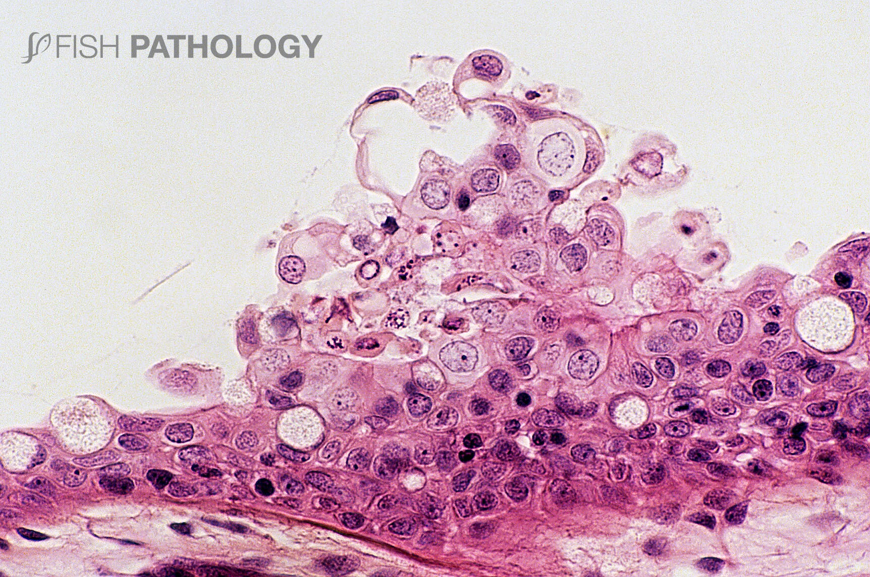
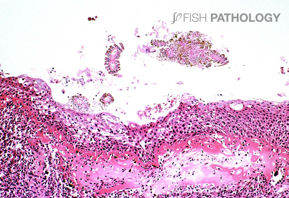
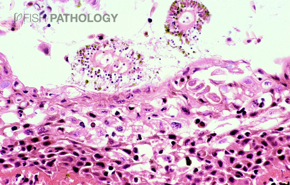
The main organs affected are the gills and skin. Changes to the respiratory epithelium involve lamellar epithelial degeneration and necrosis, exfoliation of epithelial cells, lamellar fusion, congestion, and an infiltration of inflammatory cells, leading to overall lamellar thickening.

On the arch epithelium, superficial lesions are characterized by multifocal acute ballooning degeneration, by spongiosis and hydropic degeneration, and by necrosis.

The inflammatory response is variable but is composed mainly of neutrophils. The presence of marked hyper-eosinophilia of basement membrane and the superficial subepithelial layer, suggest collagen denaturation (necrobiosis). In some cases, the gill rakers are severely necrotic, with sloughing of epithelium, and severe acute inflammation.

Figure 7. Scanning electron micrograph of Phialella quadrata showing a tentacle. It is at the tip of these tentacles that the nematocysts are found.


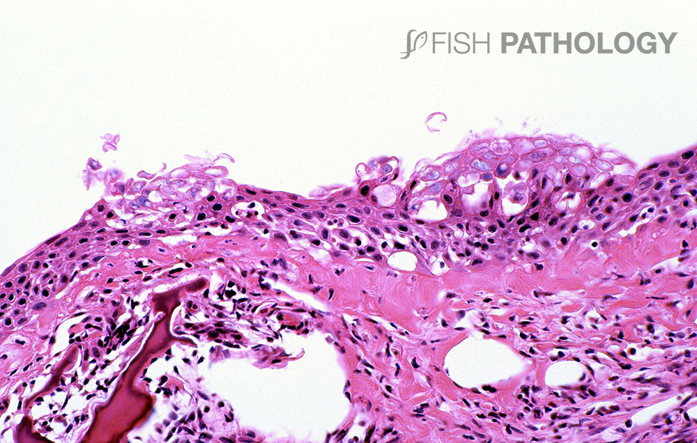
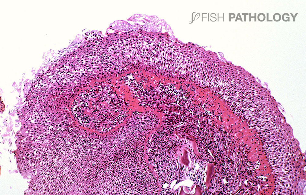
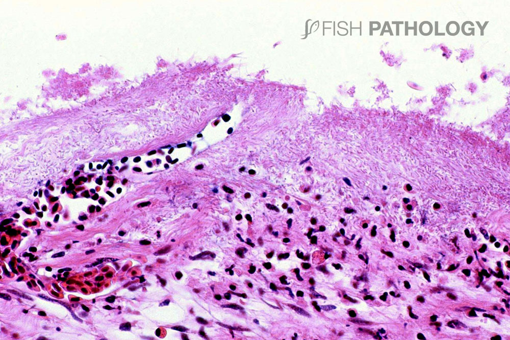
The skin lesions reveal a significant acute dermatitis characterized by a predominantly neutrophilic infiltrate, with the presence of pustule-like aggregates. The dermal stratum spongiosum is usually more affected than the stratum compactum, and haemorrhage, tissue necrosis and oedema can be observed. Generally, the underlying skeletal muscle is not affected. 


REFERENCES

  • Baxter, E. J., Sturt, M. M., Ruane, N. M., Doyle, T. K., McAllen, R., Harman, L., & Rodger, H. D. (2011). Gill damage to Atlantic salmon (Salmo salar) caused by the common jellyfish (Aurelia aurita) under experimental challenge. PLoS One6(4).
  • Baxter, E. J., Rodger, H. D., McAllen, R., & Doyle, T. K. (2011). Gill disorders in marine-farmed salmon: investigating the role of hydrozoan jellyfish. Aquaculture Environment Interactions1(3), 245-257.
  • Ferguson, H. W., Christian, M. D., Hay, S., Nicolson, J., Sutherland, D., & Crumlish, M. (2010). Jellyfish as vectors of bacterial disease for farmed salmon (Salmo salar). Journal of veterinary diagnostic investigation22(3), 376-382.
  • Marcos-López, M., Mitchell, S. O., & Rodger, H. D. (2016). Pathology and mortality associated with the mauve stinger jellyfish Pelagia noctiluca in farmed Atlantic salmon Salmo salar L. J. Fish Dis39, 111-115.
  • Purcell, J. E., Baxter, E. J., & Fuentes, V. L. (2013). Jellyfish as products and problems of aquaculture. In Advances in aquaculture hatchery technology (pp. 404-430). Woodhead Publishing.

By: Hugh Ferguson

Dr Ferguson earned his veterinary degree from the Royal (Dick) School of Veterinary Studies, Edinburgh, Scotland and held a Wellcome Research Fellowship at the Institute of Aquaculture, Stirling University where he obtained his PhD. He then worked for 4 years as a diagnostic pathologist at the Veterinary Research Laboratories, Belfast, Northern Ireland, prior to moving to Canada. He left Ontario Veterinary College after 19 years as a full professor of veterinary pathology, to return to Scotland to become head of diagnostic pathology in Stirling. During all this time he became board-certified in the American college of veterinary pathology (ACVP), and a Fellow of the Royal College of Pathologists (FRCPath, London). After Scotland he moved to become chair of veterinary pathology, and Senior Research Fellow in Windward Islands Research and Education Foundation (WINDREF), St George’s University (SGU), Grenada, West Indies. He has published more than 230 papers in refereed journals.

totop