
Nephrocalcinosis (or urolithiasis) in fish is a chronic inflammatory condition of unknown aetiology in which calcium and other minerals precipitate as hydroxyapatite within the distal renal tubules and collecting ducts.

The disease usually records low mortality and although food conversion efficiency is probably impaired, the major concern about the condition centres round a reduction in carcase quality at slaughter. In severe cases, the muscle dorsal to the kidney may also be affected.
There are some predisposing factors for this condition like high levels of carbon dioxide in water (greater than 10-20 mg/L), magnesium deficiency, selenium toxicity and a diet low in minerals. It has also been reported that chronic arsenic exposure in the diet (14mg arsenic/g) causes nephrocalcinosis in Rainbow trout.
Other factors including overcrowding and low water flow may also accentuate nephrocalcinosis; similar to high carbon dioxide levels, these lead to acidosis.
Fish with nephrocalcinosis may appear normal or show signs of abdominal swelling, variable exophthalmia, skin petechiae and haemorrhage particularly at the base of the fins.
Some fish can show an increase in skin pigmentation and the anus may protrude. Internally, ascites, splenomegaly, and inflammation of the stomach can be recorded.

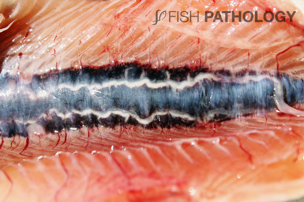
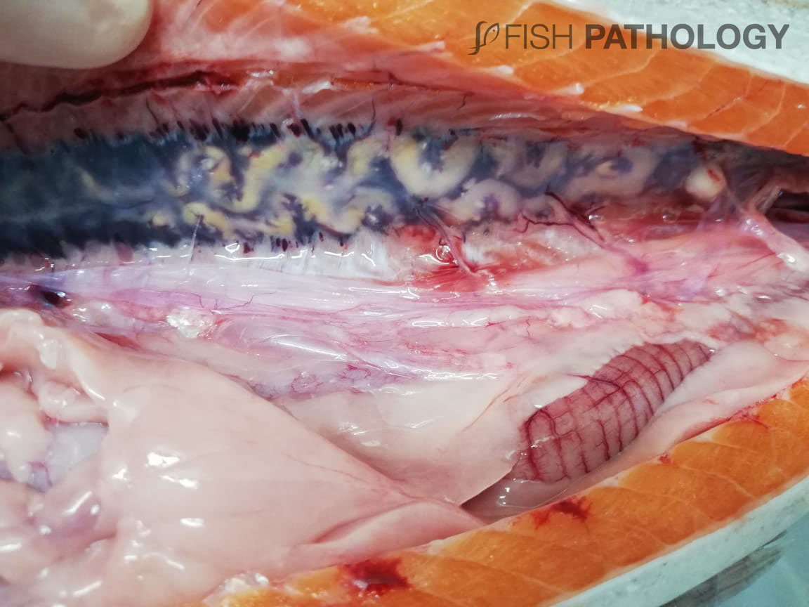
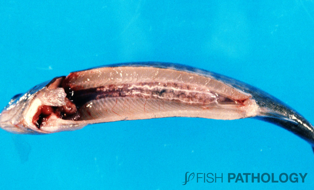
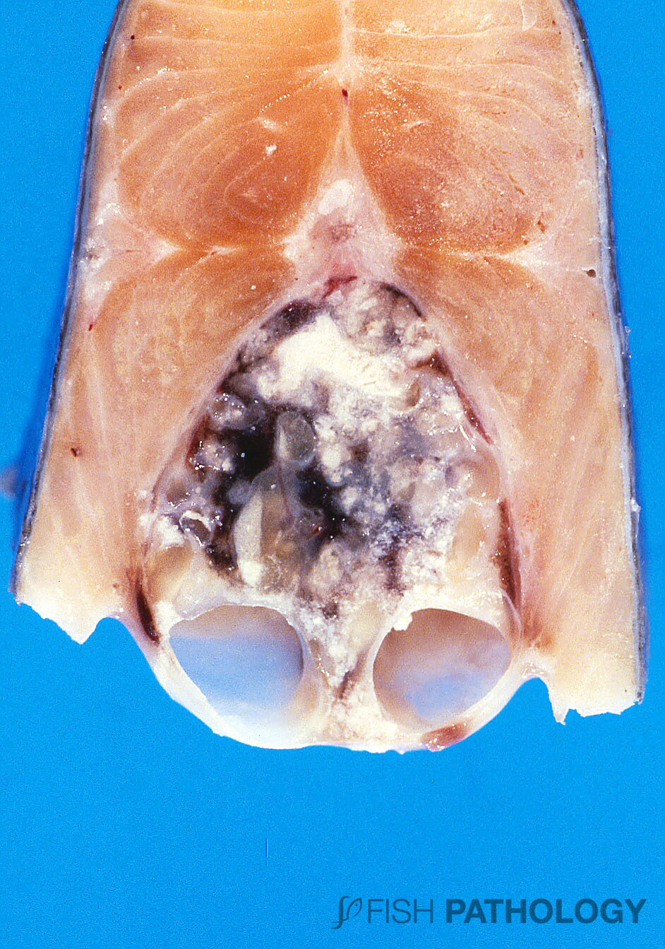
As the condition progresses the kidney becomes swollen and grey with an irregular surface and the ureters become much more sinuous as well as thickened due to the mineral deposits within.
The grossly swollen grey kidneys in nephrocalcinosis can sometimes be confused with the chronic renal inflammation seen in diseases such as bacterial kidney disease (BKD), proliferative kidney disease (PKD) or indeed some of the mycotic infections such as Exophiala. Histopathological examination quickly eliminates these differentials.
REFERENCES
- Bruno D.W.(1996). Nephrocalcinosis. Aquaculture Information Series, Marine Laboratory, Aberdeen, 16:1-5.
- Ferguson, H.W., 2006, Systemic Pathology of Fish, London, UK, Scotian Press.
- Harrison, J. G., & Richards, R. H. (1979). The pathology and histopathology of nephrocalcinosis in rainbow trout Salmo gairdneri Richardson in fresh water. Journal of Fish Diseases, 2(1), 1-12.
- Klosterhoff, M. D. C., Virginia Fonseca, P., Sampaio, L. A., Ramos, L. R. V., Tesser, M. B., & Romano, L. A. (2015). Nephrocalcinosis and kidney stones in Rachycentron canadum. Bulletin of the European Association of Fish Pathologists. 35. 138-147.
- Mousavi, S. M., Rezaie, A., Ahmadmoradi, E., & Mohammadi, F. (2016). Histopathology of nephrocalcinosis in some ornamental fishes. Aquaculture, Aquarium, Conservation & Legislation, 9(3), 574-579.
- Roberts, R.J., 2012, Fish Pathology, Oxford, USA, Blackwell Publishing Ltd.
- Cockell, K. A., Hilton, J. W., & Bettger, W. J. (1991). Chronic toxicity of dietary disodium arsenate heptahydrate to juvenile rainbow trout (Oncorhynchus mykiss). Archives of environmental contamination and toxicology, 21(4), 518-527.

1 Comments