CMS in ventricle of Atlantic salmon showing severe myocarditis of spongy layer. Note that the compact layer (on the left) is largely unaffected.

Cardiomyopathy syndrome in Atlantic salmon (CMS) – Histopathology

Cardiomyopathy syndrome (CMS) is a severe cardiac disease affecting Atlantic salmon characterized by prolonged periods of usually low-level mortalities.

Figure 1. Atrium of Atlantic salmon with CMS. Note the severity of the inflammatory response. Lesions here are often more severe than those in the ventricle. It is important, therefore, to ensure that the fragile atrium is not lost at sampling during necropsy, or in subsequent processing.

The disease was first recognized in farmed Atlantic salmon in Norway in 1985 and subsequently in farmed salmon in the Faroe Islands, Scotland and Ireland. CMS is a transmissible disease that has been causally linked to the Piscine Myocarditis Virus (PMCV), closely allied to the Totiviridae.

CMS in ventricle of Atlantic salmon showing severe myocarditis of spongy layer. Note that the compact layer (on the left) is largely unaffected.
Figure 2. CMS in ventricle of Atlantic salmon showing severe myocarditis of spongy layer. Note that the compact layer (on the left) is largely unaffected.


CMS usually causes mortality in adult or maturing fish after 12 to 18 months in seawater, resulting in substantial economic losses.

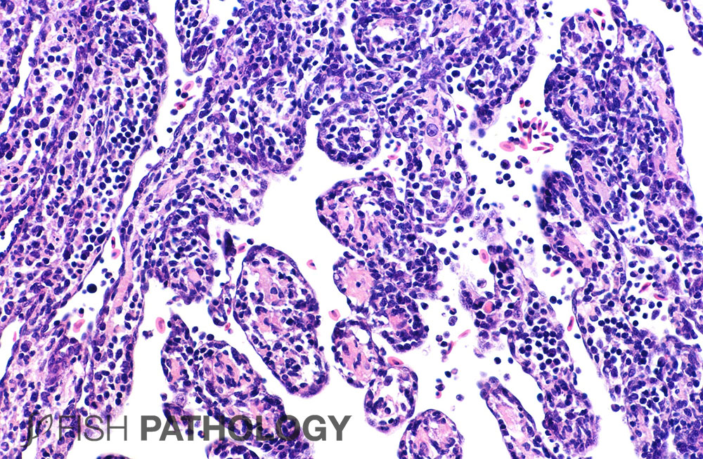
The lesions of CMS start with swollen endocardial cells, an influx of inflammatory cells into the myocardium, accompanied by degeneration, loss of striations and necrosis.

Figure 3. Later-stage CMS showing an endocardial-lined tube largely empty of muscle, but containing a few mononuclear inflammatory cells, including debris-filled macrophages, seen here with yellow cytoplasm (arrow).

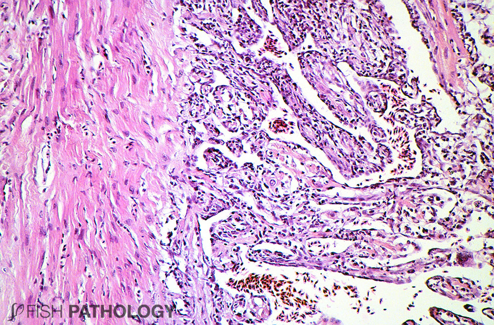
Ongoing phagocytosis of necrotic tissue leads to largely empty myocardial “tubes” filled with inflammatory cells (mostly mononuclear) including macrophages clearing up the debris. In severe cases there is frequently infiltration of lymphocyte-like cells onto the pericardium.

Figure 4. Fish in the repair stage of CMS showing a clump of myocyte-like nuclei in the centre of a cell (arrow). This appearance suggests regeneration, a process that mammals are unable to accomplish! Note the enlarged nuclei (arrow heads) on either side of this nuclear clump, possibly indicating compensatory hypertrophy of still-viable cardiac myocytes.


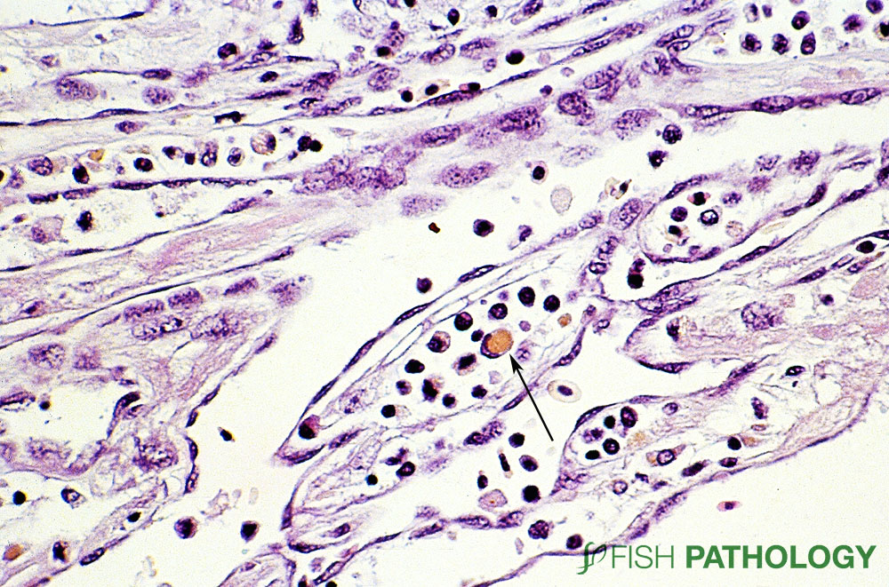
In later stages, ongoing degeneration can often be seen in the face of compensation and regeneration of myocardium.

Figure 5. Scanning electron micrograph of heart from salmon with early-stage CMS, showing crater-like loss in integrity of plasmalemma (arrows) of the endocardium. Is this the first lesion in CMS?

Figure 6. Transmission electron micrograph of heart from salmon with early-stage CMS to show the junction between 3 myocardial cells. The cells on the left and at the bottom show still-viable myofibrils (arrow), while in the cell on the right, the myofibrils have lost all structure.


Such changes include nuclear hypertrophy of myocytes adjacent to severely affected cells. But they can also include clusters of 5-20 myocyte-like nuclei, suggesting the myocardial equivalent of “nuclear rowing” as can be seen in regenerating skeletal muscle.

Liver lesions are typical of “heart failure liver” with zonal degeneration of hepatocytes, a consequence of hypoxia.


REFERENCES

  • Ferguson, H.W., T. Poppe, and D.J. Speare.  (1990). Cardiomyopathy syndrome in farmed Norwegian salmon Salmo salar L.  Dis. Aquat. Orgs. 8:225-231.
  • Garseth, Å. H., Fritsvold, C., Svendsen, J. C., Bang Jensen, B., & Mikalsen, A. B. (2018). Cardiomyopathy syndrome in Atlantic salmon Salmo salar L.: a review of the current state of knowledge. Journal of fish diseases41(1), 11-26.
  • Løvoll, M., Wiik-Nielsen, J., Grove, S., Wiik-Nielsen, C. R., Kristoffersen, A. B., Faller, R., … & Meyerson, M. (2010). A novel totivirus and piscine reovirus (PRV) in Atlantic salmon (Salmo salar) with cardiomyopathy syndrome (CMS). Virology Journal7(1), 309.
  • Wiik‐Nielsen, C. R., Ski, P. M., Aunsmo, A., & Løvoll, M. (2012). Prevalence of viral RNA from piscine reovirus and piscine myocarditis virus in Atlantic salmon, Salmo salar L., broodfish and progeny. Journal of Fish Diseases35(2), 169-171.
totop