Transmission electron micrograph from liver of fingerling tilapia with SHT. Virus particles, typical of orthomyxovirus, can be seen between and within cells (arrows).

Syncytial Hepatitis of Tilapia – Transmission Electron Microscopy

Tilapia are considered to be relatively resistant to many of the common diseases that beset other farmed fish. Viral diseases, however, are not common, and there are only a few reports in the literature.

Transmission electron micrograph from liver of fingerling tilapia with SHT. Virus particles, typical of orthomyxovirus, can be seen between and within cells (arrows).
Figure 1. Transmission electron micrograph from liver of fingerling tilapia with SHT. Virus particles, typical of orthomyxovirus, can be seen between and within cells (arrows).


Syncytial Hepatitis of Tilapia (SHT) is a newly described viral disease reported from several countries where tilapia farming is present. This virus is a member of the Orthomyxoviridae family. It has been demonstrated within hepatocytes of affected fish.


The described ultrastructural changes provide further evidence to support the previous suggestion of a viral etiology for SHT.

Transmission electron micrograph from liver of fingerling tilapia with SHT. Virus particles, typical of orthomyxovirus, can be seen between and within cells (arrows).
Figure 2. Transmission electron micrograph from liver of fingerling tilapia with SHT. Virus particles, typical of orthomyxovirus, can be seen between and within cells (arrows).


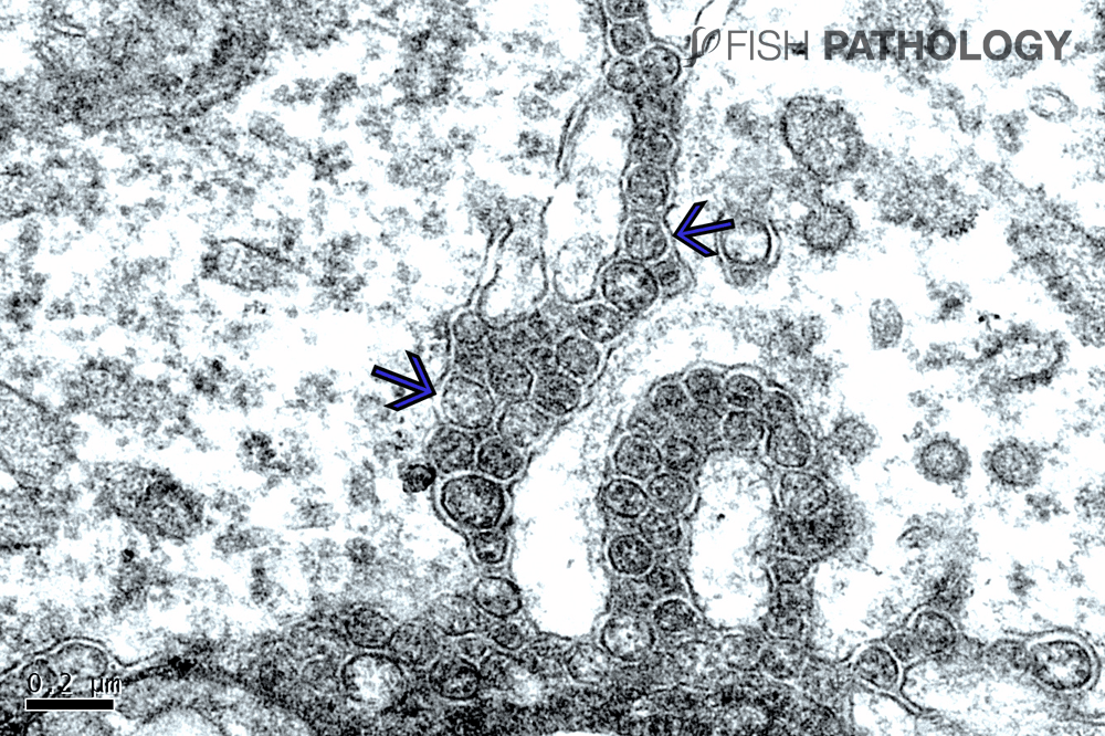
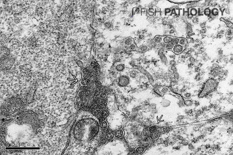
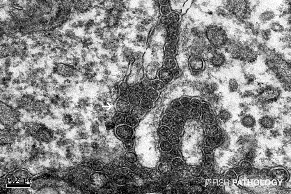
Ultrastructurally, in the liver, virions are noted only within hepatocytes and in the space of Disse and not within the endothelium. This suggests tropism of the SHT virus for the epithelial but not endothelial cell population in the liver.


When clearly visible, 60-100nm round virions with a trilaminar capsid can be seen to contain up to 7 electron-dense aggregates.

Transmission electron micrograph from liver of fingerling tilapia with SHT. Virus particles, typical of orthomyxovirus, can be seen between and within cells (arrows).
Figure 3. Transmission electron micrograph from liver of fingerling tilapia with SHT. Virus particles, typical of orthomyxovirus, can be seen between and within cells (arrows).


REFERENCES

  • Del-Pozo, J., Mishra, N., Kabuusu, R., Cheetham, S., Eldar, A., Bacharach, E., … & Ferguson, H. W. (2017). Syncytial hepatitis of tilapia (Oreochromis niloticus L.) is associated with orthomyxovirus-like virions in hepatocytes. Veterinary pathology54(1), 164-170.
  • Ferguson, H. W., Kabuusu, R., Beltran, S., Reyes, E., Lince, J. A., & Del Pozo, J. (2014). Syncytial hepatitis of farmed tilapia, Oreochromis niloticus (L.): a case report. J Fish Dis37(6), 583-589.

By: Hugh Ferguson

Dr Ferguson earned his veterinary degree from the Royal (Dick) School of Veterinary Studies, Edinburgh, Scotland and held a Wellcome Research Fellowship at the Institute of Aquaculture, Stirling University where he obtained his PhD. He then worked for 4 years as a diagnostic pathologist at the Veterinary Research Laboratories, Belfast, Northern Ireland, prior to moving to Canada. He left Ontario Veterinary College after 19 years as a full professor of veterinary pathology, to return to Scotland to become head of diagnostic pathology in Stirling. During all this time he became board-certified in the American college of veterinary pathology (ACVP), and a Fellow of the Royal College of Pathologists (FRCPath, London). After Scotland he moved to become chair of veterinary pathology, and Senior Research Fellow in Windward Islands Research and Education Foundation (WINDREF), St George’s University (SGU), Grenada, West Indies. He has published more than 230 papers in refereed journals.

totop