Liver; O. niloticus L.; dissociation of hepatocytes containing brown-staining lipoprotein, degeneration and single cell necrosis, plus early hepatocellular syncytial cell formation and moderate diffuse inflammation H&E.

Syncytial Hepatitis of Tilapia – Histopathology

Syncytial Hepatitis of Tilapia (SHT) is a newly described viral disease reported from Ecuador, Israel, Colombia and several other countries where tilapia farming is present.

Liver; O. niloticus L.; dissociation of hepatocytes containing brown-staining lipoprotein, degeneration and single cell necrosis, plus early hepatocellular syncytial cell formation and moderate diffuse inflammation H&E.
Figure 1. Liver; O. niloticus L.; dissociation of hepatocytes containing brown-staining lipoprotein, degeneration and single cell necrosis, plus early hepatocellular syncytial cell formation and moderate diffuse inflammation H&E.

The most consistently described target organs are the liver and gastrointestinal tract, although there are some reports of lesions within the central nervous system.


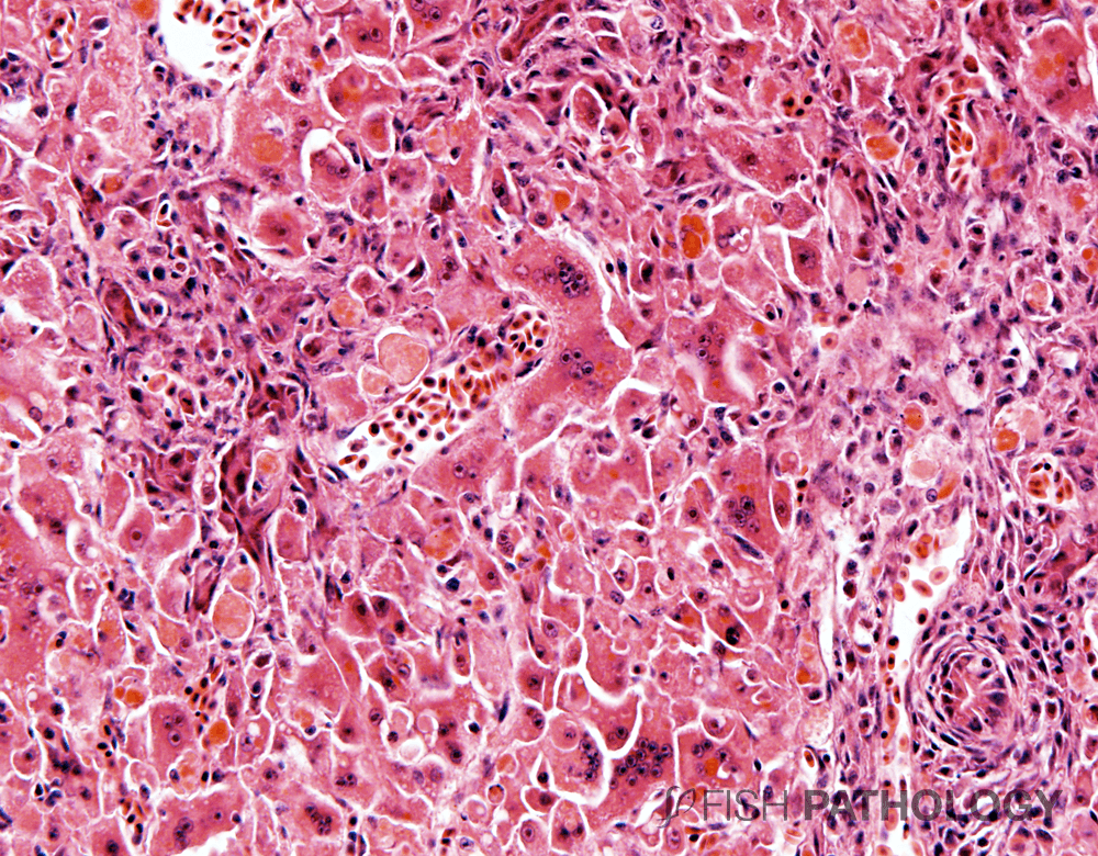
The presence of a novel virus with close affinity to the family Orthomyxoviridae has been demonstrated within hepatocytes of affected fish.


Histopathological lesions include necrotizing hepatitis with distinctive hepatocellular syncytial giant cell formation, dissociation of hepatocytes often with accumulation of lipoprotein, a predominantly lymphocytic inflammatory infiltration.

Stomach from fingerling tilapia showing acute gastritis, especially on the right of the image. Here we can also see loss of gastric glands. H&E.
Figure 2. Stomach from fingerling tilapia showing acute gastritis, especially on the right of the image. Here we can also see loss of gastric glands. H&E.


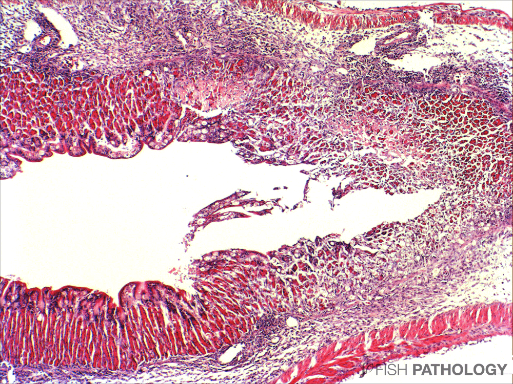
In the gastrointestinal tract, necrosis of gastric glandular epithelium and intestinal enterocytes can be observed together with protein casts, as can loss of pancreatic acini.


REFERENCES

  • Del-Pozo, J., Mishra, N., Kabuusu, R., Cheetham, S., Eldar, A., Bacharach, E., … & Ferguson, H. W. (2017). Syncytial hepatitis of tilapia (Oreochromis niloticus L.) is associated with orthomyxovirus-like virions in hepatocytes. Veterinary pathology54(1), 164-170.
  • Ferguson, H. W., Kabuusu, R., Beltran, S., Reyes, E., Lince, J. A., & Del Pozo, J. (2014). Syncytial hepatitis of farmed tilapia, Oreochromis niloticus (L.): a case report. J Fish Dis37(6), 583-589.

By: Hugh Ferguson

Dr Ferguson earned his veterinary degree from the Royal (Dick) School of Veterinary Studies, Edinburgh, Scotland and held a Wellcome Research Fellowship at the Institute of Aquaculture, Stirling University where he obtained his PhD. He then worked for 4 years as a diagnostic pathologist at the Veterinary Research Laboratories, Belfast, Northern Ireland, prior to moving to Canada. He left Ontario Veterinary College after 19 years as a full professor of veterinary pathology, to return to Scotland to become head of diagnostic pathology in Stirling. During all this time he became board-certified in the American college of veterinary pathology (ACVP), and a Fellow of the Royal College of Pathologists (FRCPath, London). After Scotland he moved to become chair of veterinary pathology, and Senior Research Fellow in Windward Islands Research and Education Foundation (WINDREF), St George’s University (SGU), Grenada, West Indies. He has published more than 230 papers in refereed journals.

totop